Irene Margaret Vavasour1, Jackie T Yik2,3, Pierre Becquart4, Jasmine Gill4, Shannon H Kolind1,2,3,5, Alice J Schabas5, Ana-Luiza Sayao5, Virginia Devonshire5, Robert Carruthers5, Anthony Traboulsee5, GR Wayne Moore3,4,5, Sophie Stukas4, Cheryl Wellington4, Jacqueline Quandt4, David KB Li1, and Cornelia Laule1,2,3,4
1Radiology, University of British Columbia, Vancouver, BC, Canada, 2Physics and Astronomy, University of British Columbia, Vancouver, BC, Canada, 3International Collaboration on Repair Discoveries (ICORD), University of British Columbia, Vancouver, BC, Canada, 4Pathology and Laboratory Medicine, University of British Columbia, Vancouver, BC, Canada, 5Medicine, University of British Columbia, Vancouver, BC, Canada
1Radiology, University of British Columbia, Vancouver, BC, Canada, 2Physics and Astronomy, University of British Columbia, Vancouver, BC, Canada, 3International Collaboration on Repair Discoveries (ICORD), University of British Columbia, Vancouver, BC, Canada, 4Pathology and Laboratory Medicine, University of British Columbia, Vancouver, BC, Canada, 5Medicine, University of British Columbia, Vancouver, BC, Canada
Diffusely abnormal white matter (DAWM) was
found in 35% of clinically isolated syndrome (CIS) and ~60% of multiple sclerosis
participants. CIS with DAWM had more negative brain health markers (e.g. smaller
cortical thickness, more lesions, higher neurofilament) compared to CIS without
DAWM.
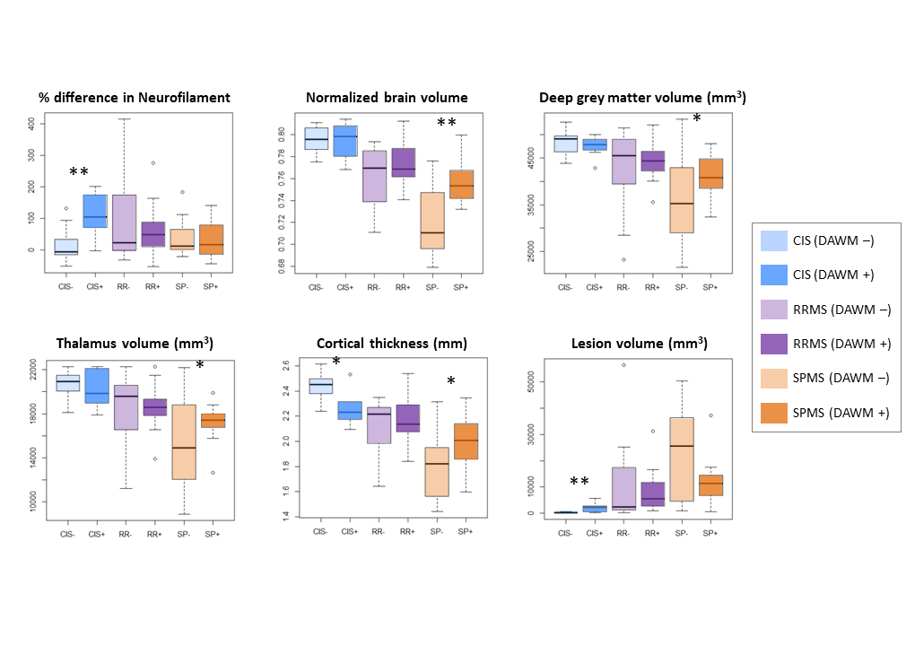
Figure 4: Boxplots divided based on
participant subtype (clinically isolated syndrome (CIS), relapsing-remitting
multiple sclerosis (RR)
and
secondary progressive multiple sclerosis (SP)) with (+) and without (–) diffusely
abnormal white matter
(DAWM). Neurofilament
values were normalised to matched healthy control values. Significance between
DAWM– and
DAWM+ are indicated with *p<0.05, **p<0.005.
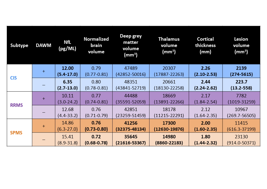
Figure 3: Neurofilament (NfL) and MRI
measurements (mean
and (range)) for the
different MS subtypes with (+) and without (–) diffusely abnormal white matter
(DAWM) (CIS: clinically isolated syndrome; RRMS: relapsing-remitting multiple
sclerosis; SPMS: secondary progressive multiple). Bolded values indicate a significant
difference between DAWM+ and
DAWM–.