Gian Franco Piredda1,2,3, Tom Hilbert1,2,3, Manuela Vaneckova4, Jan Krasensky4, Michaela Andelova5, Tomas Uher5, Barbora Srpova5, Eva Kubala Havrdova5, Karolina Vodehnalova5, Dana Horakova5, Veronica Ravano1,2,3, Bénédicte Maréchal1,2,3, Jean-Philippe Thiran2,3, and Tobias Kober1,2,3
1Advanced Clinical Imaging Technology, Siemens Healthcare AG, Lausanne, Switzerland, 2Department of Radiology, Lausanne University Hospital and University of Lausanne, Lausanne, Switzerland, 3LTS5, École Polytechnique Fédérale de Lausanne (EPFL), Lausanne, Switzerland, 4Department of Radiology, First Faculty of Medicine, Charles University and General University Hospital, Prague, Poland, 5Department of Neurology and Center of Clinical Neuroscience, First Faculty of Medicine, Charles University and General University Hospital, Prague, Poland
1Advanced Clinical Imaging Technology, Siemens Healthcare AG, Lausanne, Switzerland, 2Department of Radiology, Lausanne University Hospital and University of Lausanne, Lausanne, Switzerland, 3LTS5, École Polytechnique Fédérale de Lausanne (EPFL), Lausanne, Switzerland, 4Department of Radiology, First Faculty of Medicine, Charles University and General University Hospital, Prague, Poland, 5Department of Neurology and Center of Clinical Neuroscience, First Faculty of Medicine, Charles University and General University Hospital, Prague, Poland
Using high-resolution, whole brain T1 mapping, a periventricular gradient of abnormality was detected in normal-appearing white matter, increasing from early to progressive stages of multiple sclerosis.
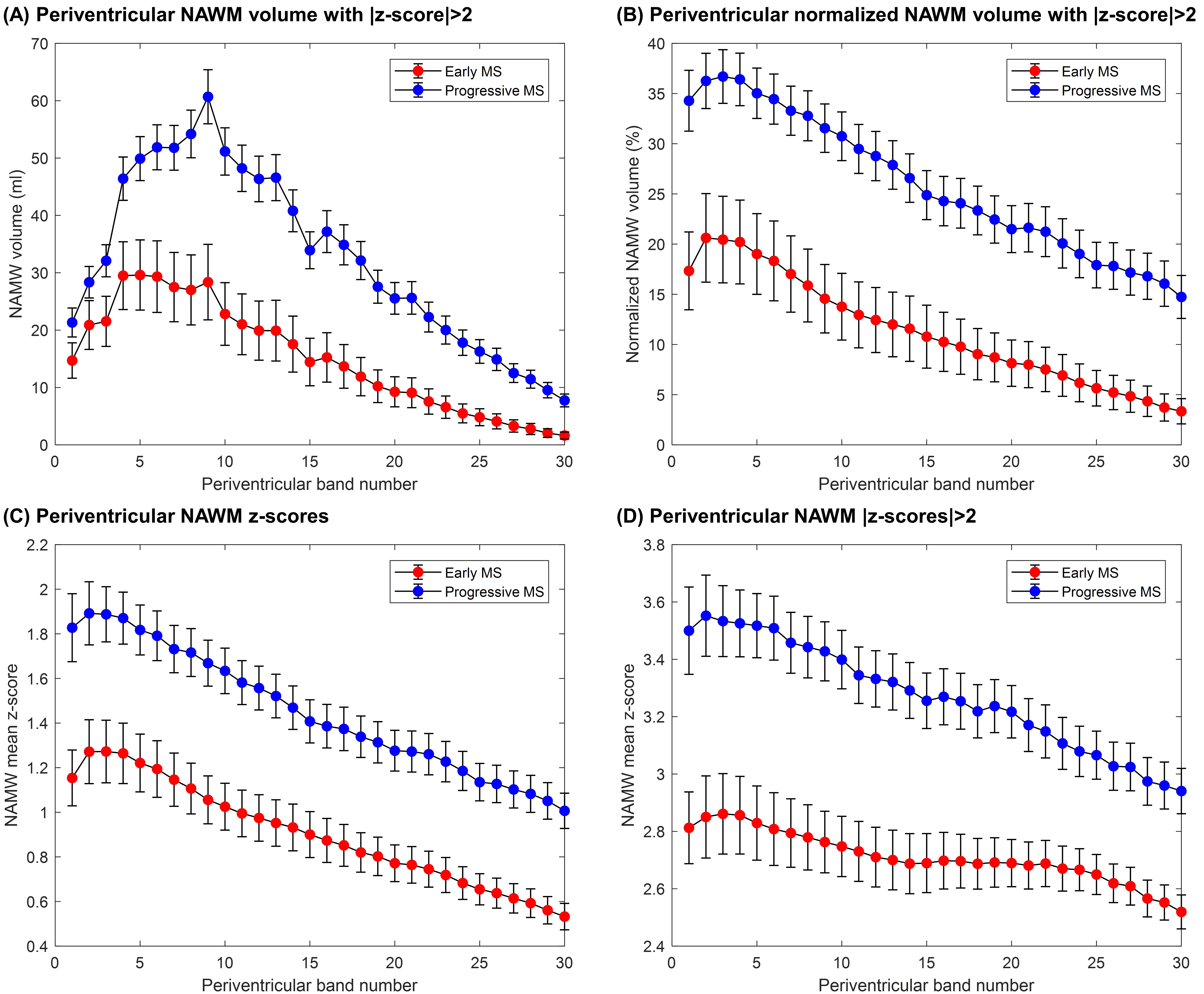
Figure 4. Periventricular gradients of (A) abnormal WM volume (ml) and (B) normalized abnormal WM volume to the total band volume, which were identified as the NAWM voxel exceeding a z-score threshold of 2 (i.e. exceeding the prediction interval at 95% level of confidence of the T1 normative linear model). Periventricular gradients in NAWM of (C) absolute z-scores and (D) absolute z-scores > 2. Error bars indicate two standard errors. Reported values in the first band should be carefully considered as they may be affected by partial-volume effects between CSF and WM tissues.
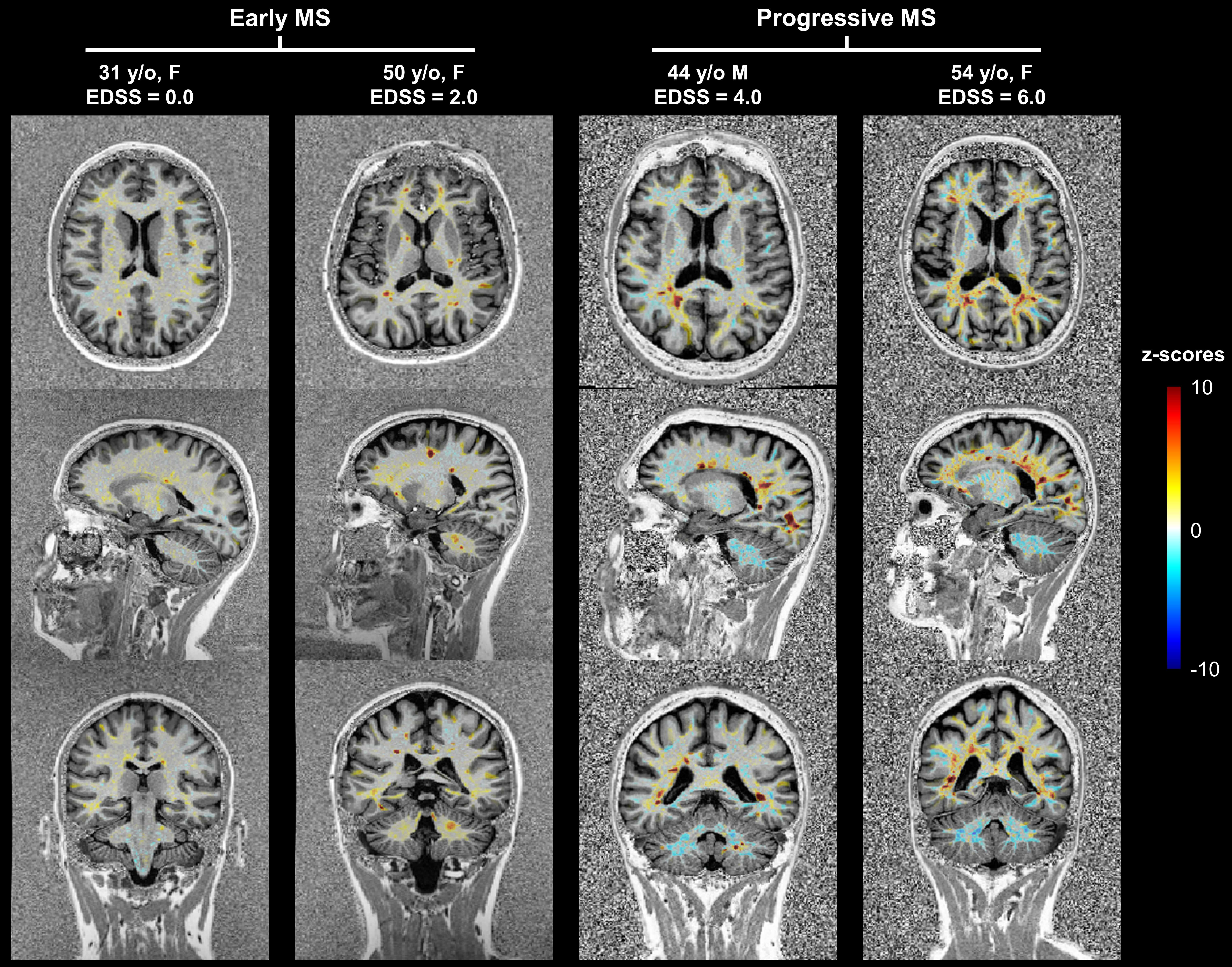
Figure 2. Representative example z-score maps overlaid onto the MP2RAGE contrast in early MS patients with low Expanded Disability Status Scale (EDSS) scores and progressive MS patients with higher EDSS.