Jason Michael Millward1, Claudia Chien2, Joseph Kuchling2, Friedemann Paul2, Thoralf Niendorf1, and Sonia Waiczies1
1Berlin Ultrahigh Field Facility (B.U.F.F.), Max Delbrück Center for Molecular Medicine in the Helmholtz Association, Berlin, Germany, 2NeuroCure Clinical Research Center, Charité - Universitätsmedizin Berlin, Berlin, Germany
1Berlin Ultrahigh Field Facility (B.U.F.F.), Max Delbrück Center for Molecular Medicine in the Helmholtz Association, Berlin, Germany, 2NeuroCure Clinical Research Center, Charité - Universitätsmedizin Berlin, Berlin, Germany
Longitudinal 3T scans of patients with
clinically isolated syndrome reveal that brain ventricle volumes do not exclusively
expand unidirectionally but in some patients can expand and contract, even
over a period of years. Patients with contracting ventricles were younger than
those without.
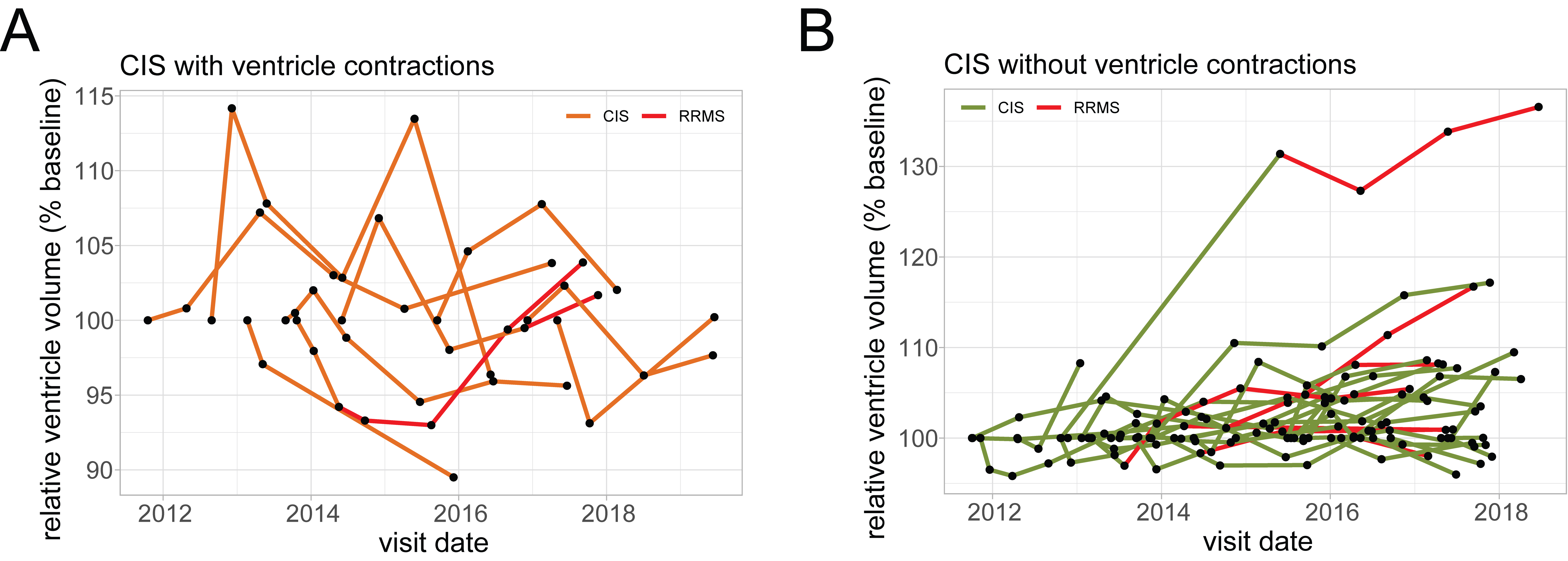
Relative ventricle volumes for individual patients (conversion to RRMS
depicted by red). A. A substantial
fraction of the CIS patients (23%) showed contractions of ventricle volumes
over time, beyond the range of variation in healthy subjects (±6%). B. The majority of CIS patients did not
show contractions in ventricle volume beyond the range of variation of healthy
subjects. Many of these patients, both those who converted to RRMS and those
who did not, showed increased ventricle volume over time, consistent with brain
atrophy and neurodegeneration.
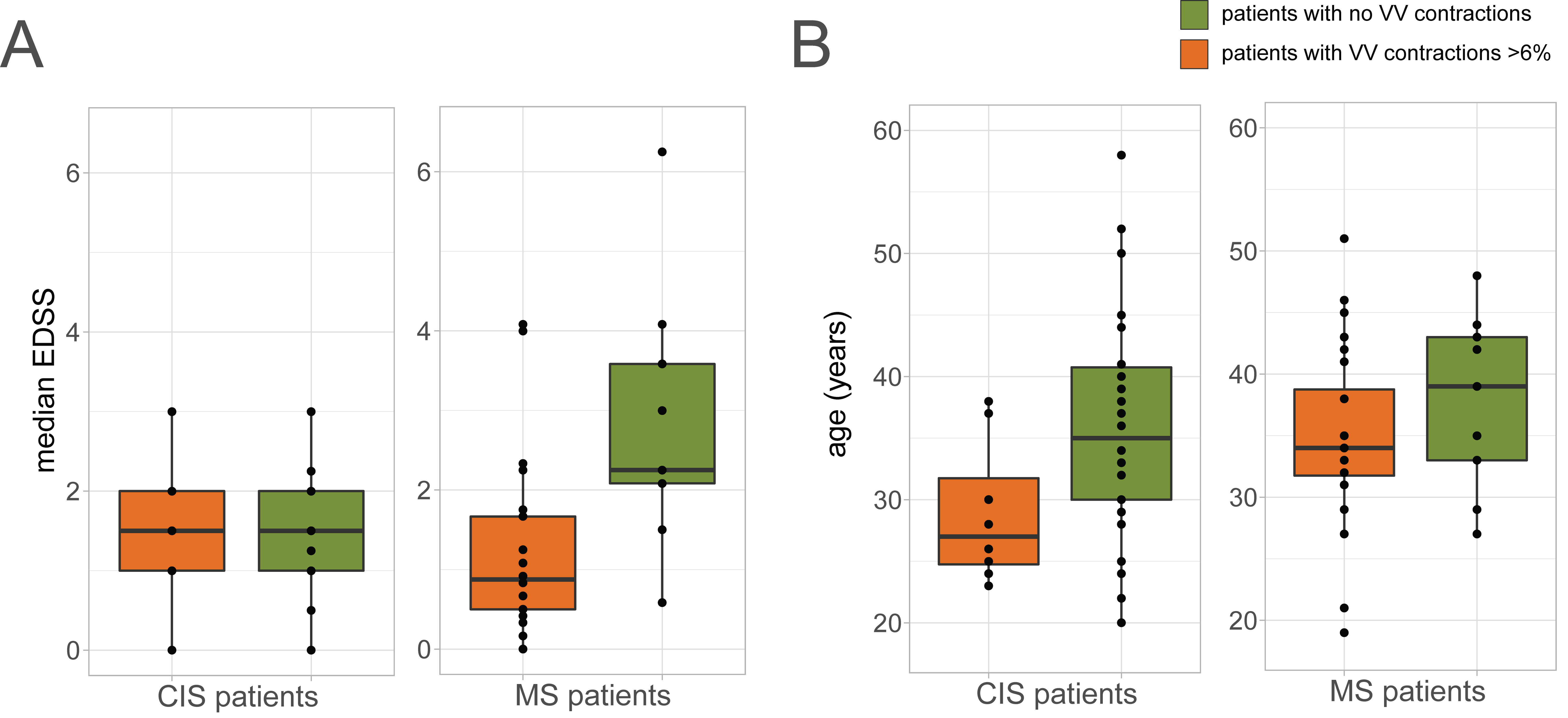
A. There was no
significant difference in median EDSS between CIS patients with and without
ventricle contractions >6%. The EDSS of the MS patients with contractions was
similar to that of the CIS patients; EDSS of MS patients with contractions was
significantly lower than that of MS patients without contractions (p=0.0063,
Mann-Whitney test). B. CIS patients
with ventricle contractions >6% were significantly younger than those
without (p=0.0447, Mann-Whitney test). In the MS cohort, patients with
ventricle contractions trended lower in age, though this is not significant.
