Kyle Jeong1, You-Jung Lee1, Suk-Keu Yeom2, Noel Carlson3, Lubdha Shah4, John Rose5, and Eun-Kee Jeong4
1Utah Center for Advanced Imaging research, University of Utah, Salt Lake City, UT, United States, 2Radiology, Korea University Ansan Hospital, Ansan, Korea, Republic of, 3GRECC, Veteran Affairs, Salt Lake City, UT, United States, 4Radiology and Imaging Sciences, University of Utah, Salt Lake City, UT, United States, 5Neuroimmunology Division, University of Utah, Salt Lake City, UT, United States
1Utah Center for Advanced Imaging research, University of Utah, Salt Lake City, UT, United States, 2Radiology, Korea University Ansan Hospital, Ansan, Korea, Republic of, 3GRECC, Veteran Affairs, Salt Lake City, UT, United States, 4Radiology and Imaging Sciences, University of Utah, Salt Lake City, UT, United States, 5Neuroimmunology Division, University of Utah, Salt Lake City, UT, United States
The purpose of the study was to perform a quantitative evaluation of myelination on genetically engineered myelin-deficit shiverer mice and wild-type mice using ultrahigh-b diffusion MRI (UHb-DWI).
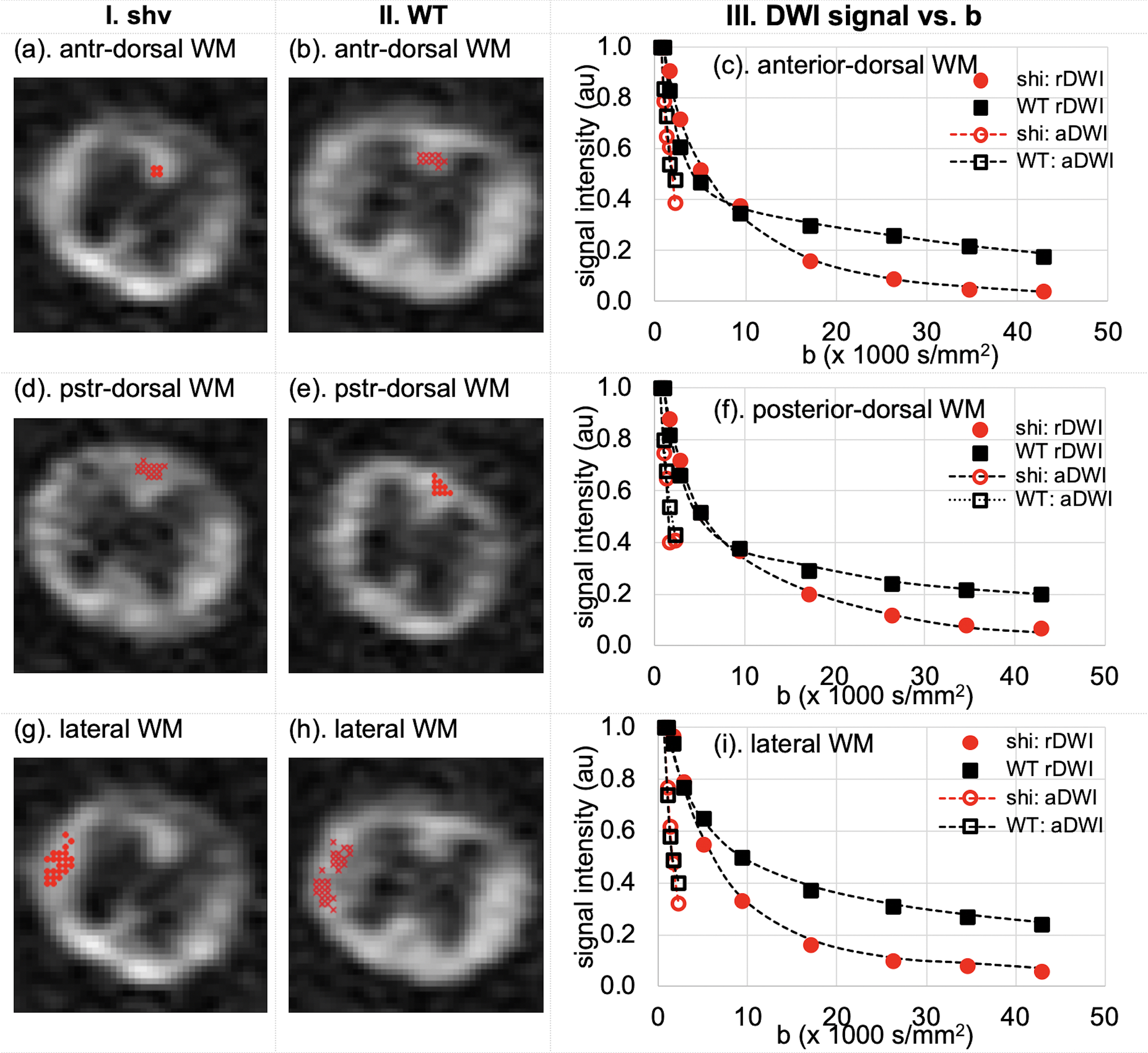
Fig. 1. DWIs of b=9380 s/mm2 of (col I) shiverer and (col II) WT mice spinal cords, and signal-b curves of (c) anterior-dorsal and (f) posterior-dorsal column, and (i) lateral white-matter tracts up to b = 42,890 s/mm2 for UHb-rDWI and 2210 s/mm2 for UHb-aDWI. Signals were averaged over two nearby slices of two shiverer (red) and two adjacent slices of six WT (black) mouse spinal cords.
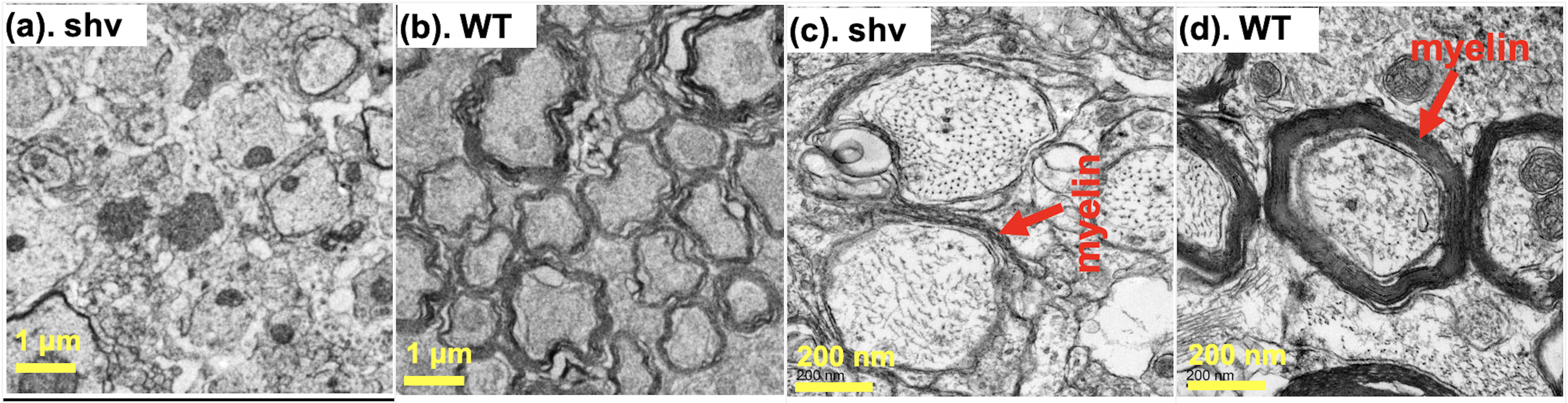
Fig. 2. EM images of dorsal (sensory) column of (a, c) shiverer and (b, d) WT mice spinal cords in two different magnifications (a, b) x1,100 and (c, d) x11,000. In this EM picture, the myelin sheaths of the shiverer mouse is less numbered and looser than that of the wild-type mouse.