Marios C. Yiannakas1, Ratthaporn Boonsuth1, Carmen Tur1,2, Marco Battiston1, Francesco Grussu1,3, Rebecca S. Samson1, Torben Schneider4, Masami Yoneyama5, Ferran Prados1,6,7, Sara Collorone1, Rosanna Cortese1, Olga Ciccarelli1, and Claudia A. M. Gandini Wheeler-Kingshott1,8,9
1NMR Research Unit, Queen Square MS Centre, Department of Neuroinflammation, UCL Queen Square Institute of Neurology, Fuculty of Brain Sciences, University College London, London, United Kingdom, 2Multiple Sclerosis Centre of Catalonia (Cemcat), Vall d’Hebron Institute of Research, Vall d'Hebron Barcelona Hospital Campus, Barcelona, Spain, 3Radiomics Group, Vall d’Hebron Institute of Oncology, Vall d’Hebron Barcelona Hospital Campus, Barcelona, Spain, 4Philips Healthcare, Surrey, Guildford, United Kingdom, 5Philips Japan, Minatoku, Tokyo, Japan, 6Centre for Medical Image Computing, Medical Physics and Biomedical Engineering, University College London, London, United Kingdom, 7Universitat Oberta de Catalunya, Barcelona, Spain, 8Department of Brain and Behavioural Sciences, University of Pavia, Pavia, Italy, 9Brain Connectivity Research Centre, IRCCS Mondino Foundation, Pavia, Italy
1NMR Research Unit, Queen Square MS Centre, Department of Neuroinflammation, UCL Queen Square Institute of Neurology, Fuculty of Brain Sciences, University College London, London, United Kingdom, 2Multiple Sclerosis Centre of Catalonia (Cemcat), Vall d’Hebron Institute of Research, Vall d'Hebron Barcelona Hospital Campus, Barcelona, Spain, 3Radiomics Group, Vall d’Hebron Institute of Oncology, Vall d’Hebron Barcelona Hospital Campus, Barcelona, Spain, 4Philips Healthcare, Surrey, Guildford, United Kingdom, 5Philips Japan, Minatoku, Tokyo, Japan, 6Centre for Medical Image Computing, Medical Physics and Biomedical Engineering, University College London, London, United Kingdom, 7Universitat Oberta de Catalunya, Barcelona, Spain, 8Department of Brain and Behavioural Sciences, University of Pavia, Pavia, Italy, 9Brain Connectivity Research Centre, IRCCS Mondino Foundation, Pavia, Italy
In this pilot in vivo study, magnetisation transfer
ratio measurements in the sciatic nerve of people with relapsing-remitting multiple
sclerosis show significantly reduced values as compared to healthy controls
suggesting alteration in myelin content.
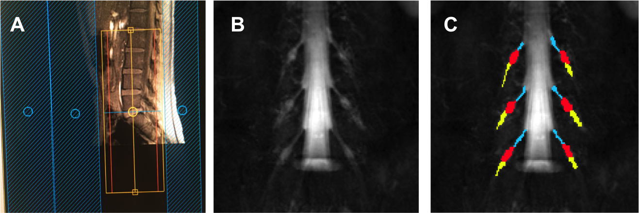
Figure 1. A) Prescription
of the 3D SHINKEI and MTR sequences for imaging the lumbar plexus in the
coronal plane: B) Example image obtained using the 3D SHINKEI at the level of
the lumbar plexus (L2-L4 segments shown); C) Manual segmentation
of the lumbar segments with separate binary masks created for the
preganglionic, ganglionic and postganglionic regions shown in blue, red,
yellow, respectively.
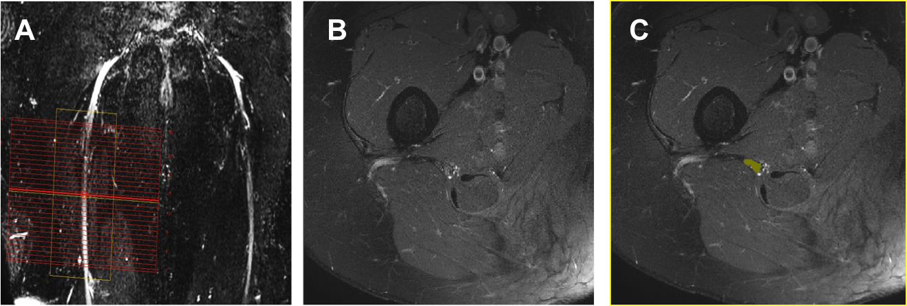
Figure 2. A) Prescription
of the high-resolution
fat-suppressed T2-weighted and MTR sequences for imaging the sciatic
nerve in the axial plane; B) Example image obtained using the high-resolution
fat-suppressed T2-weighted sequence; C) Manual segmentation of the sciatic
nerve (binary mask shown in yellow).