Fedel Machado-Rivas1,2, Camilo Jaimes1,2, Benoit Scherrer1,2, Mark Gorman1,2, Simon K Warfield1,2, and Onur Afacan1,2
1Radiology, Boston Children's Hospital, Boston, MA, United States, 2Radiology, Harvard Medical School, Boston, MA, United States
1Radiology, Boston Children's Hospital, Boston, MA, United States, 2Radiology, Harvard Medical School, Boston, MA, United States
In addition to finding differences in cAD, cRD, cMD and cFA, compartment diffusion model heterogeneity index was found to be significantly different in lesions. Our results support that a DIAMOND analysis can provide insights to MS lesion microstructure beyond conventional DTI metrics.
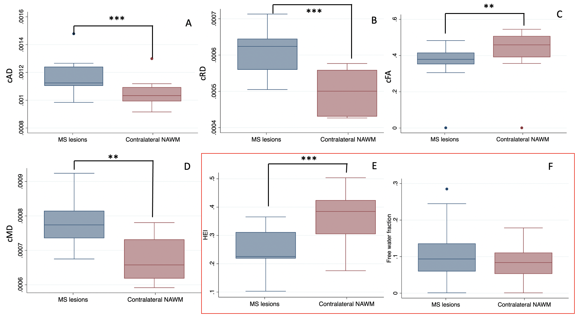
Comparison of compartment Axial Diffusivity (cAD) (A), compartment Radial Diffusivity (cRD) (B), compartment Fractional Anisotropy (cFA) (C), ompartment Mean Diffusivity (cMD) (D), heterogeneity index (HEI) (E), and free water fraction (F) between MS lesions and Contralateral NAWM in patients. Significance levels for Mann-Whitney U tests are displayed as *P<0.05, **P≤0.01, ***P≤0.001.
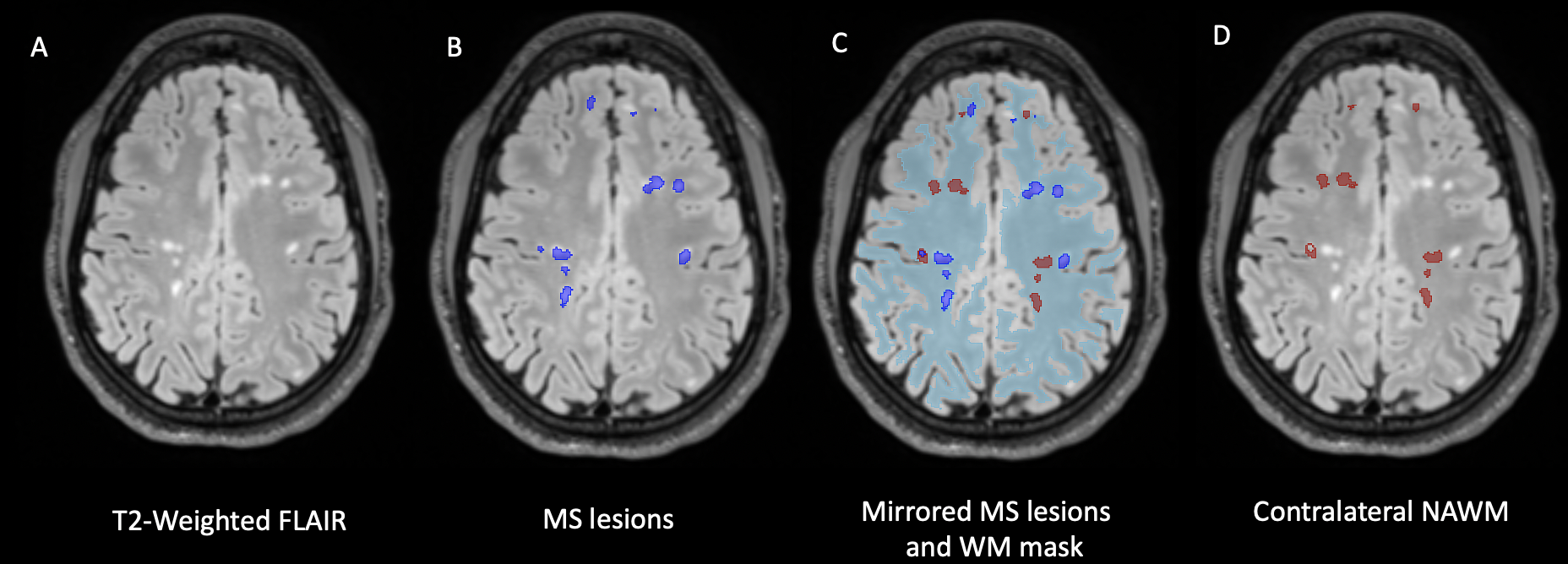
Lesion and contralateral normal appearing white matter (NAWM) segmentation in T2 weighted FLAIR acquisitions. Hyperintense white matter lesions (A) were manually segmented (B). A linear transform was used to mirror lesions in the contralateral side of the brain, and a white matter mask was used to refine segmentations (C,D).