Øystein Bech Gadmar1, Anne-Hilde Farstad2, Berit Elstad2, Piotr Sowa2, and Wibeke Nordhøy1
1Diagnostic Physics, Oslo University Hospital, Oslo, Norway, 2Radiology, Oslo University Hospital, Oslo, Norway
1Diagnostic Physics, Oslo University Hospital, Oslo, Norway, 2Radiology, Oslo University Hospital, Oslo, Norway
By modelling T2 prepared double inversion MR, a "True-T2" DIR sequence was found and tested that removes the undesired T1 contrast in fluid attenuated imaging therefore improving the desired T2 contrast between lesions both in WM and GM. Comparison with traditional FLAIR imaging was favorable.
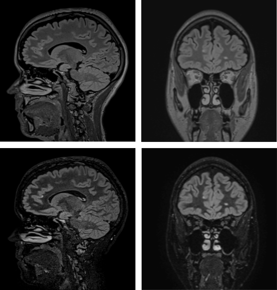
Fluid attenuated IR
images of a MS patient. Left/right: Sagittally acquired vs coronally
reformatted images. Top panels: FLAIR. Bottom panels: True-T2 DIR of
similar image planes. Multiple focal lesions are seen in the images,
situated in deep WM as well as GM-associated.
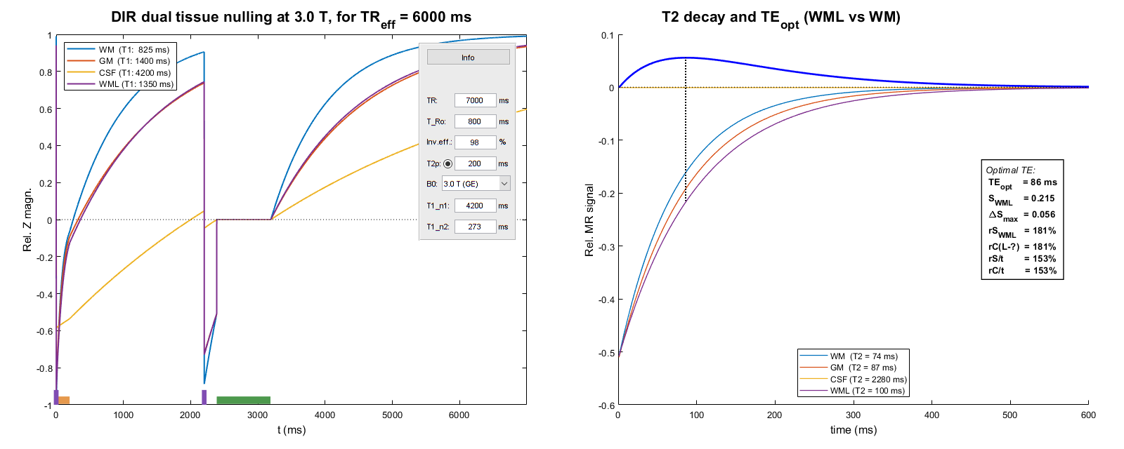
Simulated
True-T2 DIR sequence with a T2 preparation phase. Figure elements as
in fig. 1; in addition an information box showing the simulator
settings is included in the left panel. In the right panel is shown an
information box showing signal and contrast strength and efficiency,
that is, relative SNR/CNR per unit of acquisition time. The
percentages shown refer to the corresponding ones in fig. 1.