Ivan Jambor1, Aida Steiner2, Marko Pesola3, Timo Liimatainen4, Marcus Sucksdorff3, Eero Rissanen 2, Laura Airas2, Hannu Aronen3, and Harri Merisaari3
1Icahn School of Medicine at Mount Sinai, New York, NY, United States, 2Turku University Hospital, Turku, Finland, 3University of Turku, Turku, Finland, 4University of Oulu, Oulu, Finland
1Icahn School of Medicine at Mount Sinai, New York, NY, United States, 2Turku University Hospital, Turku, Finland, 3University of Turku, Turku, Finland, 4University of Oulu, Oulu, Finland
Whole brain T1ρadiab and TRAFF2 at 3T was
feasible with significant differences in T1ρadiab and TRAFF2 values between
tissues time and correlation with disease severity.
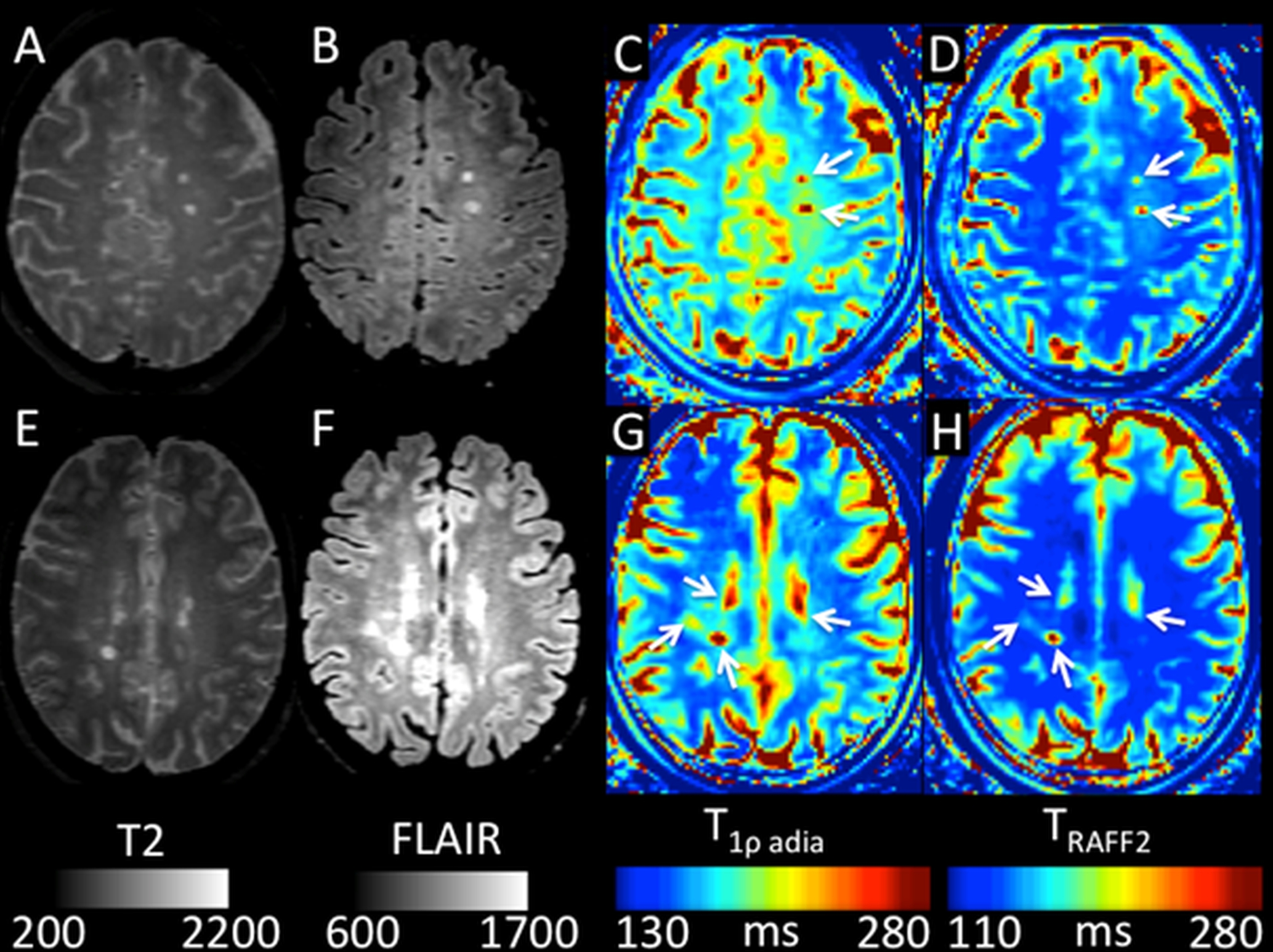
T2-weighted imaging (A, E), FLAIR
(B, F), T1radiab (C, G) and TRAFF2 (D, H)
imaging findings (white arrow) in a 47-year-old female with disease
progression, EDSS score at baseline of 2.5 and
1-year follow up of 3.0, (A, B, C, D) and 46-year-old female with no
change in her baseline EDSS score of 2.5 at 1-year follow up (E, F, G, H).
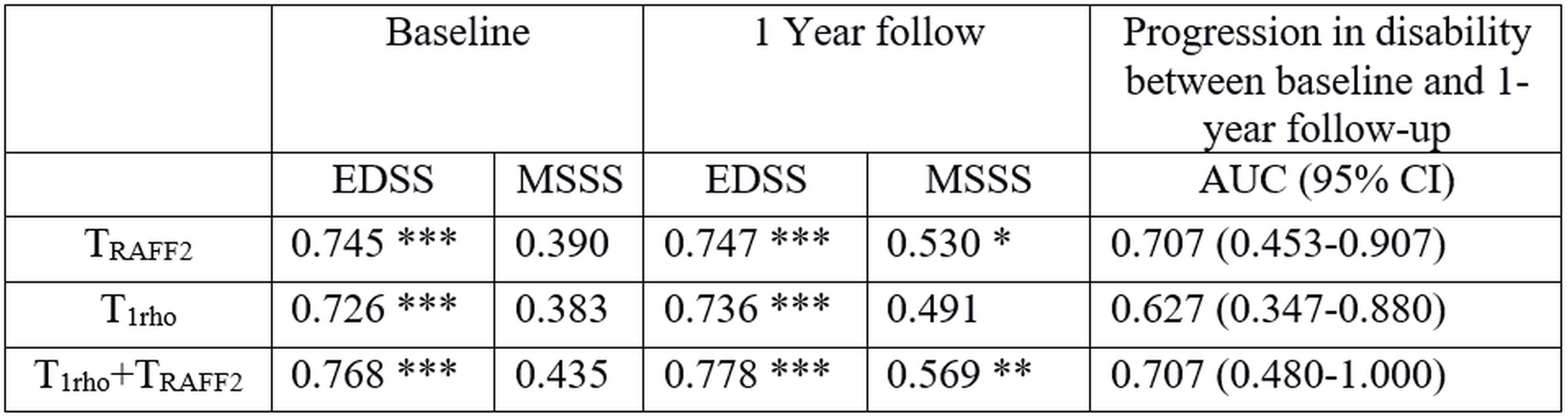
Potential of adiabatic T1rho and Relaxation Along
a Fictitious Field imaging (TRAFF2) for prediction of
Expanded Disability Status Scale (EDSS) and Multiple Sclerosis Severity Score
(MSSS), correcting for age of patients. The values are R2, with statistically significant correlations after
Bonferroni correction over 12 evaluations marked as (raw p-value threshold): *
0.05 (p<4.17x10⁻³), ** 0.01 (p<8.33 x10⁻⁴) and ***
0.001 (p<8.33 x10⁻⁵). AUC = Area Under Receiver Operating
Characteristic Curve; 95% CI = 95% Confidence Interval.