Alina Lopatina1,2, Stefan Ropele3, Renat Sibgatulin1, Jürgen R Reichenbach1,2,4, and Daniel Güllmar1
1Medical Physics Group / IDIR, Jena University Hospital, Jena, Germany, 2Michael-Stifel-Center for Data-Driven and Simulation Science, Jena, Germany, 3Department of Neurology, Medical University of Graz, Graz, Austria, 4Center of Medical Optics and Photonics Jena, Jena, Germany
1Medical Physics Group / IDIR, Jena University Hospital, Jena, Germany, 2Michael-Stifel-Center for Data-Driven and Simulation Science, Jena, Germany, 3Department of Neurology, Medical University of Graz, Graz, Austria, 4Center of Medical Optics and Photonics Jena, Jena, Germany
The
classification procedure of identifying multiple sclerosis based on
diffusion-weighted imaging by using convolutional neural networks was
analyzed by generating relevance maps. The study showed that the central brain area and some of the lesion voxels are important for
classification.
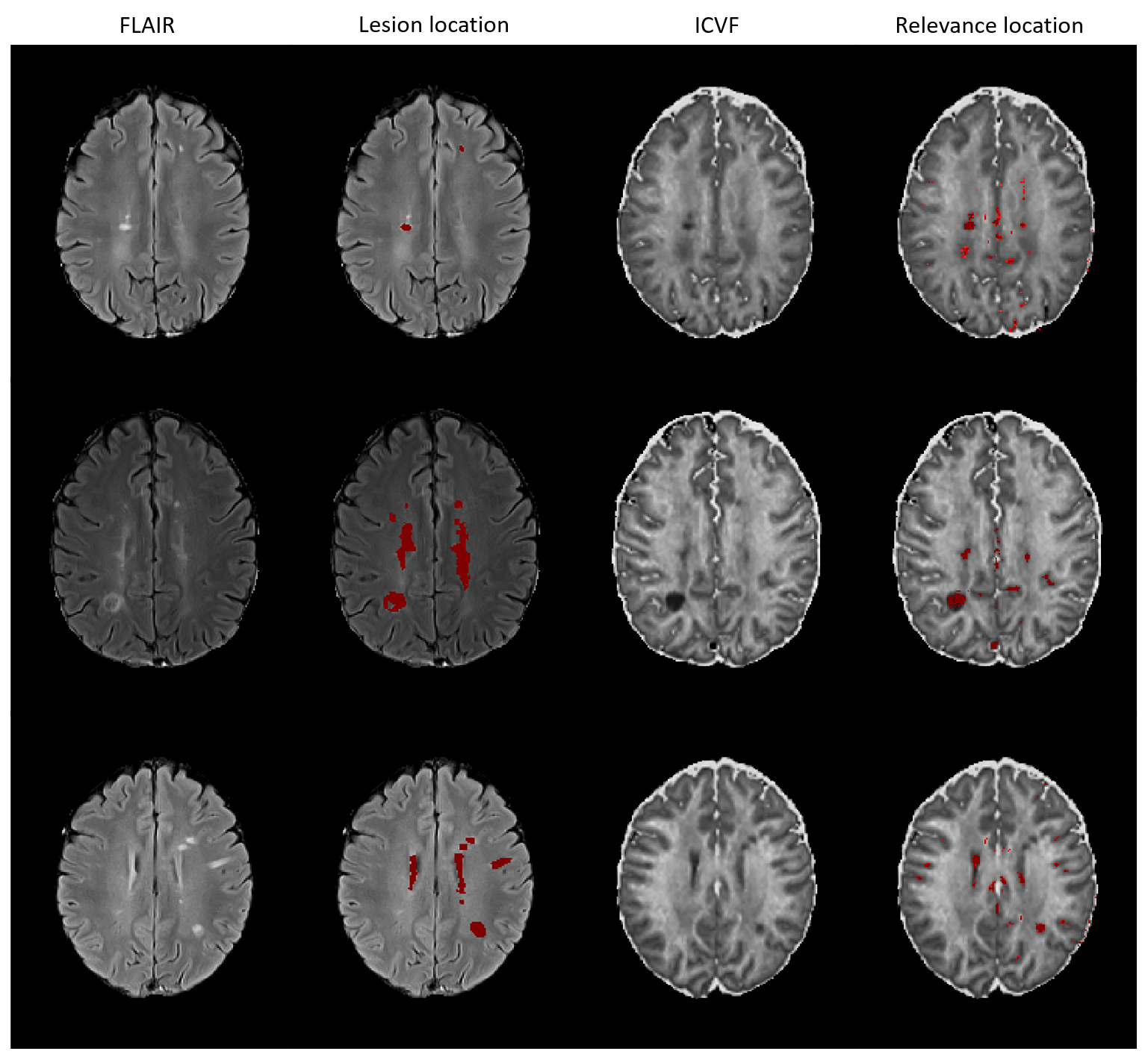
Figure
3. Comparison of FLAIR-based lesion maps and ICVF-based relevance maps for
three correctly classified MS patients (in rows). Red shows the brain areas with
lesions in the lesion maps and positive relevant voxels in relevance maps.
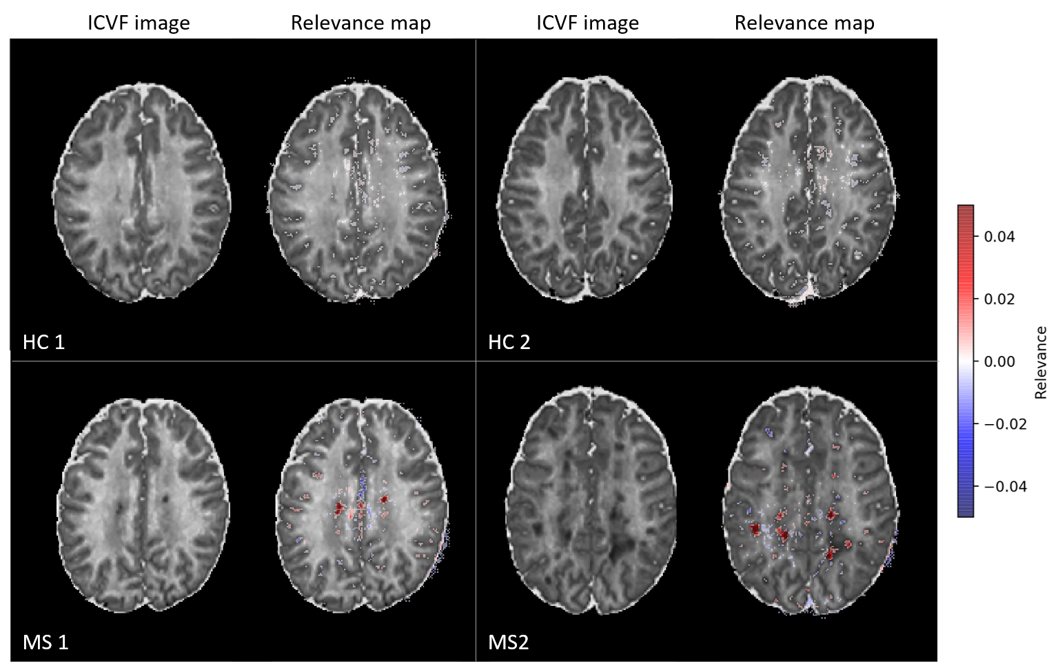
Figure
1. ICVF images and corresponding relevance maps overlaid on the corresponding ICVF
images for two correctly classified HC and two correctly classified MS
subjects. Red shows the positive relevance of the voxel information for correct
classification and blue indicates voxels possessing a negative relevance.