Stephen T. Vito1, Kai H. Barck2, Rohan S. Virgincar2, Amy Easton1, Robby M. Weimer2, Tracy J. Yuen1, and Luke Xie2
1Neuroscience, Genentech, South San Francisco, CA, United States, 2Biomedical Imaging, Genentech, South San Francisco, CA, United States
1Neuroscience, Genentech, South San Francisco, CA, United States, 2Biomedical Imaging, Genentech, South San Francisco, CA, United States
MRI detected inflammation-mediated demyelination in the cuprizone model. Voxel-based analysis revealed significant lesions in the medial splenium, lateral genu, and cerebellar nuclei. T2 reflected inflammation (microglia and astrocytes) while MWF and gRatio suggested demyelination.
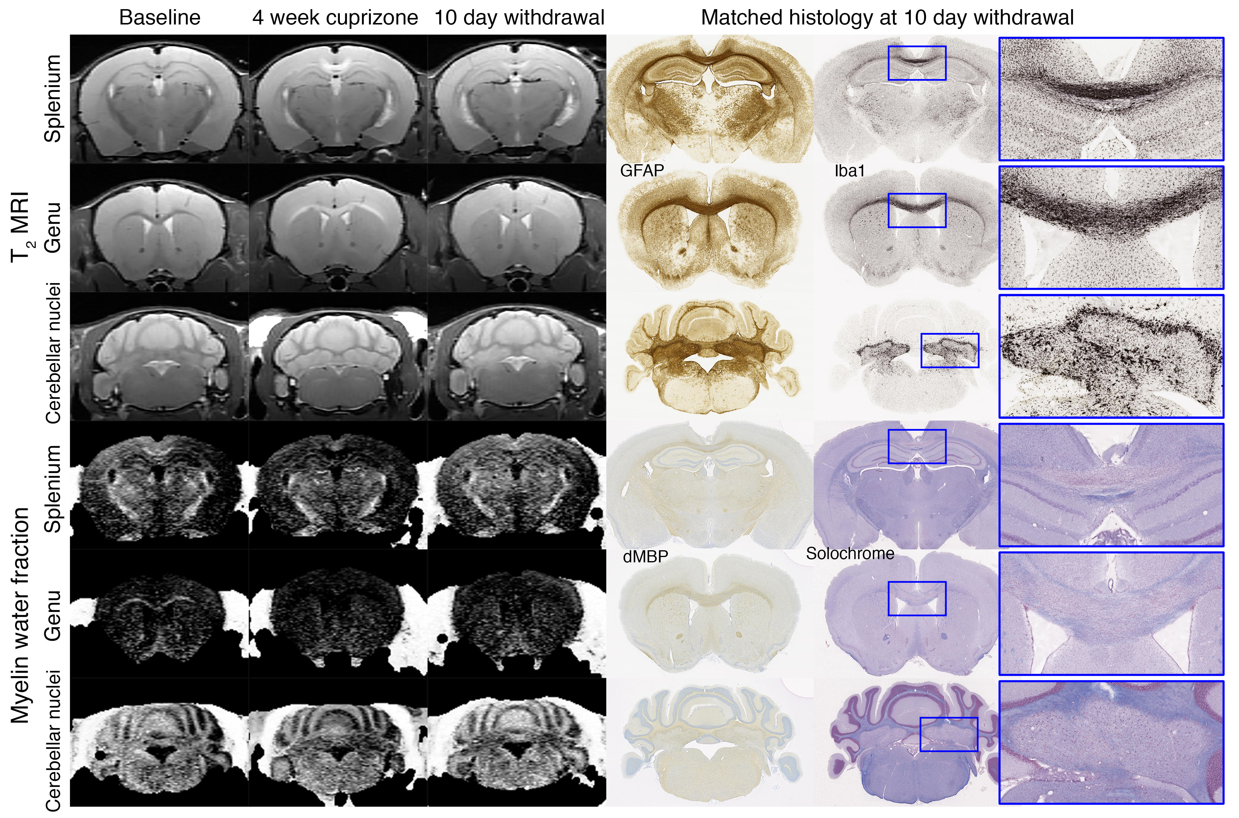
Fig. 1. Longitudinal MRI of brain lesions at baseline, 4-week cuprizone, and 10-day withdrawal (n=12). Lesions are observed in the splenium, genu, and cerebellar nuclei in T2-weighted and myelin water fraction images. Example histology of the same animals are shown. Inserts show lesion areas at 5× magnification. GFAP~astrocytes, Iba1~microglia, dMBP~denatured myelin basic protein, and solochrome~myelin.
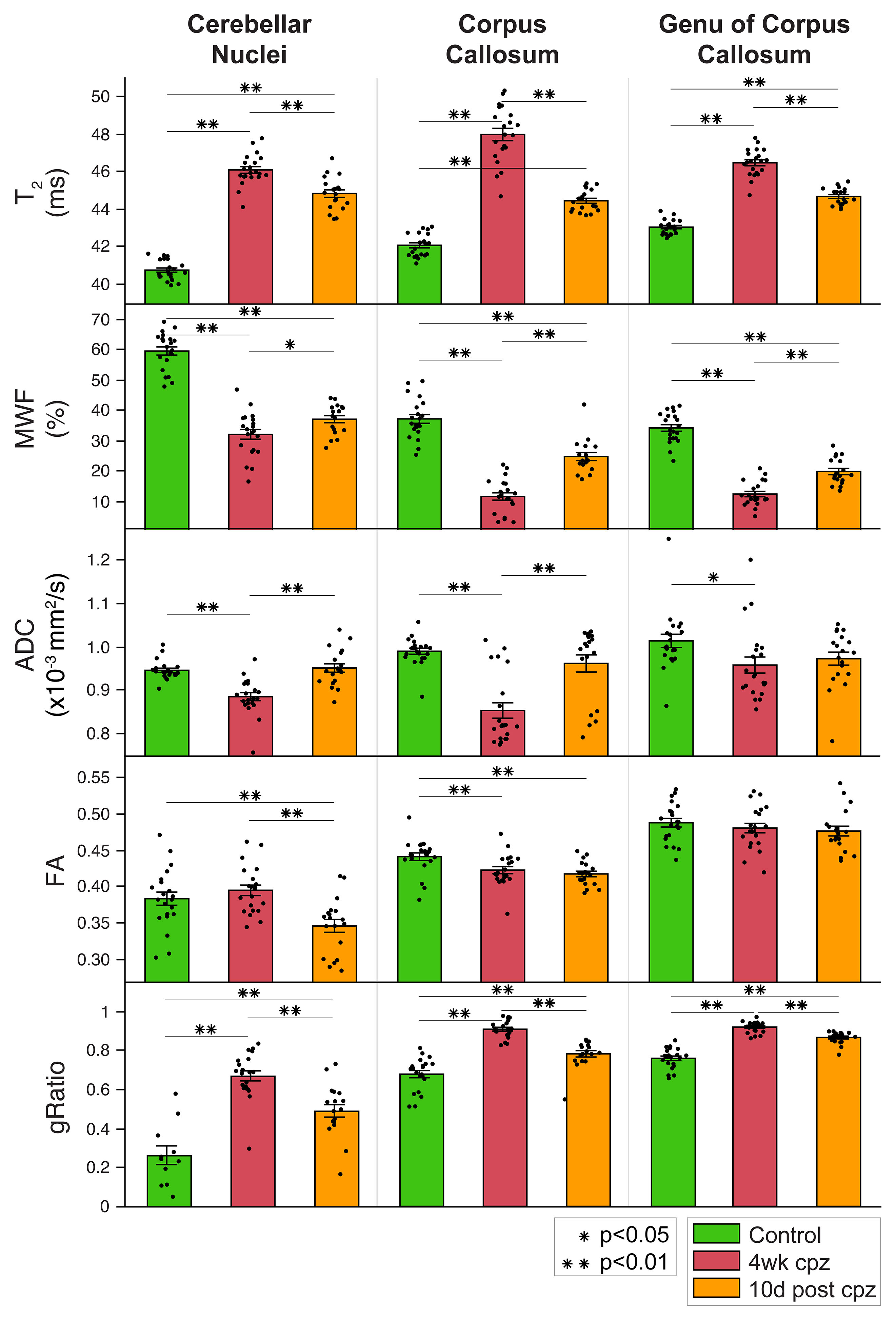
Fig. 2. Bar plots for longitudinal MRI. Error bars show standard error about the mean. Sample size: n=12 at baseline, 4-week cuprizone, and 10-day withdrawal. MWF=myelin water fraction, ADC=apparent diffusion coefficient, and FA=fractional anisotropy. One-way ANOVA and post-hoc t-tests were performed between baseline control, 4-week cuprizone, and 10-day withdrawal groups (*p<0.05, **p<0.01).