Ya-Jun Ma1, Hyungseok Jang1, Zhao Wei1, Mei Wu1, Saeed Jerban1, Eric Y Chang1,2, Jody Corey-Bloom1, Graeme M Bydder1, and Jiang Du1
1UC San Diego, San Diego, CA, United States, 2VA Health system, San Diego, CA, United States
1UC San Diego, San Diego, CA, United States, 2VA Health system, San Diego, CA, United States
UltraShort T2
Proton Fraction (USPF) reduction in multiple
sclerosis (MS) lesions
suggests that the proposed STAIR-dUTE-ES technique has potential for
evaluation of demyelination and remyelination in the diagnosis and treatment of
patients with MS.
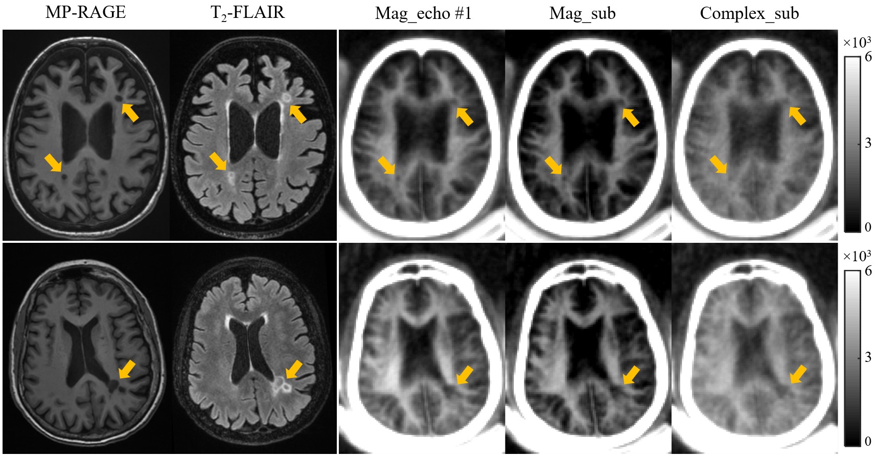
Figure 3 Selective clinical MP-RAGE (first
column), T2-FLAIR (second column) and STAIR-dUTE (last three
columns) images of two representative patients with MS (first row: a 49-year-old
female; second row: a 69-year-old female). MS lesions appeared hypointense on
the MP-RAGE image and hyperintense on the T2-FLAIR image as indicated
by the yellow arrows. These lesions also show signal loss on the magnitude images in the first echo images (third
column), magnitude echo subtracted images (fourth column) and complex echo
subtracted images (last column) using the STAIR-dUTE sequence.
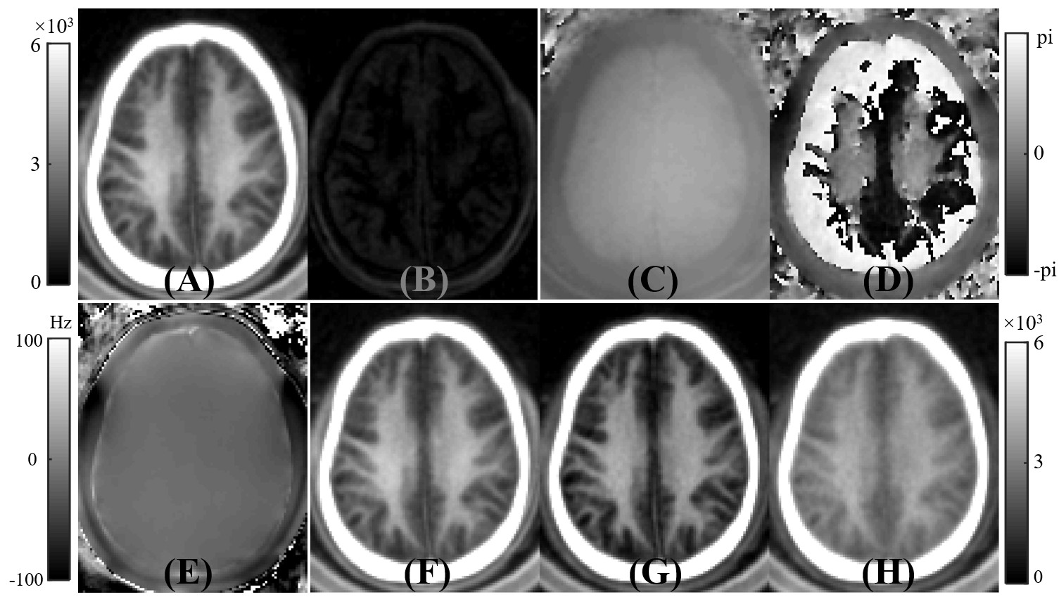
Figure 2 A volunteer study showing the generation of ultrashort T2 signals with the methods used in this study. The first row shows magnitude
images for the first echo (A) and the second echo (B), as well as the corresponding
phase images for the first echo (C) and second echo (D). Panel E shows the ΔB0
field map used for the complex ES. Magnitude of the first echo (F), magnitude
echo subtracted (G), and complex echo subtracted (H) images obtained with the STAIR-dUTE
sequence are shown. The magnitude of the first echo image (A) is displayed
again in (F) for closer comparison with (G) and (H).