1Radiological Sciences, UCLA Geffen School of Medicine, Los Angeles, CA, United States, 2School of Health Sciences, Purdue University, West Lafayette, IN, United States, 3UCLA School of Nursing, Los Angeles, CA, United States
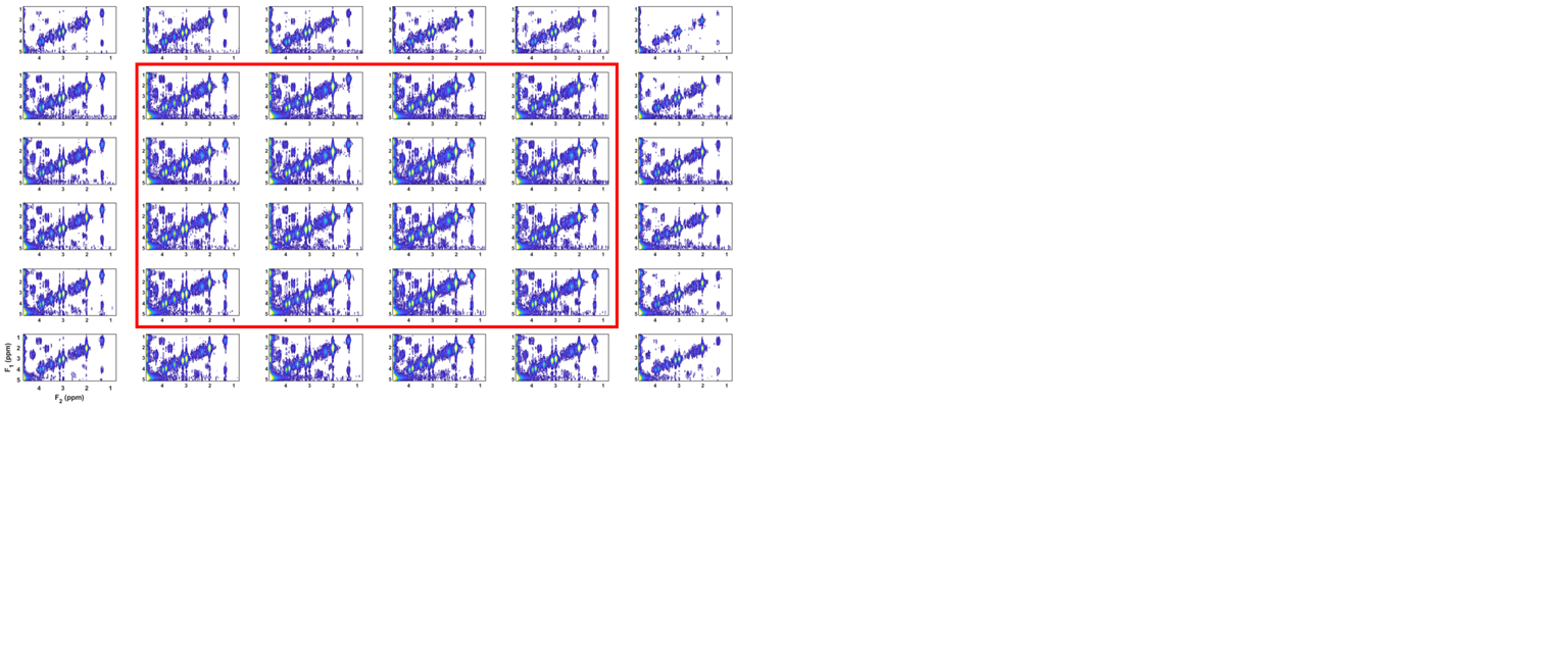
Test-retest reproducibility of a novel 5D COSI-CONCEPT sequence was validated using a GE Braino on two different 3T MRI scanners. The phantom was scanned on different days and we observed excellent coefficients of variance (CV) and intra-class correlation coefficients for all 6 metabolites
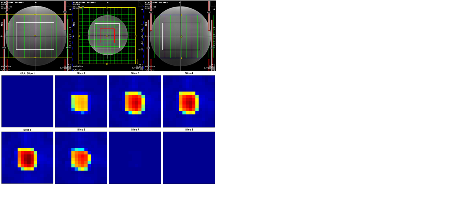
Fig. 2: (Top) Three-plane localization of the volume-of-interest (VOI) in the brain phantom. The VOI size was 10.5cm × 10.5cm × 7.5cm and the field-of-view (FOV) for spectroscopic imaging was 24cm × 24cm × 12cm along the left-to-right (L-R), anterior-to-posterior (A-P) and foot-to-head (F-H) directions, respectively. Sixteen voxels (red square) within the VOI were quantified. (Bottom) Axial NAA maps acquired along the F-H dimension within the FOV. Signal from five slices was measurable within the VOI, which had an extent of 7.5 cm along F-H.