Linda Bianchini1, João Santinha2,3, Francesca Botta4, Daniela Origgi4, Marta Cremonesi4, and Alessandro Lascialfari1
1University of Pavia, Pavia, Italy, 2Champalimaud Center for the Unknown, Lisbon, Portugal, 3Instituto Superior Técnico, Lisbon, Portugal, 4European Institute of Oncology IRCCS, Milan, Italy
1University of Pavia, Pavia, Italy, 2Champalimaud Center for the Unknown, Lisbon, Portugal, 3Instituto Superior Técnico, Lisbon, Portugal, 4European Institute of Oncology IRCCS, Milan, Italy
A study on T2-weighted MR images of a radiomic phantom mimicking pelvic tumors shows that the texture discriminative power of radiomic features depends on tumor volume, with most features losing this property below 1 cm3 at 1.5 T.
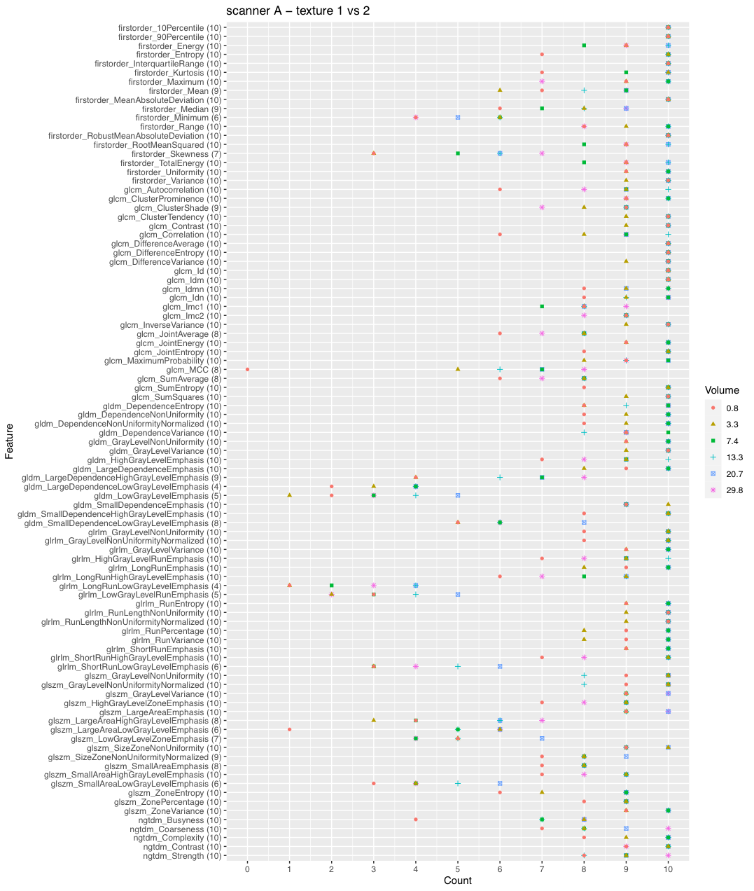
Figure 2. Discrimination of textures 1 and 2 on scanner A. The plot shows the number of times a feature is able to discriminate texture 1 and 2 if extracted from original and filtered images acquired on scanner A, as a function on the VOI size. Due to the selection of features with ICC>0.9, not all the features were extracted from all the available image types (10 in total: original + 9 filtered images), so the maximum count a feature could reach is indicated next to the feature name on y axis.
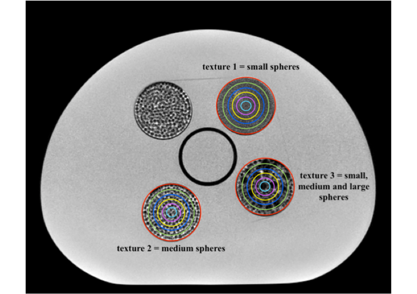
Figure 1. Identified VOIs and textures. Three phantom inserts were chosen as representative of three textures, from a finer texture (1) to a coarser one (3), through a medium texture (2). The volumes of the selected VOIs were: 29.8 cm3 (red), 20.7 cm3 (green), 13.3 cm3 (blue), 7.4 cm3 (yellow), 3.3 cm3 (pink), and 0.8 cm3 (cyan).