Mohsen Farzi1, Irvin Teh1, Darryl McClymont2, Hannah Whittington2, Craig A. Lygate2, and Jürgen E. Schneider1
1Cardiovascular & Metabolic Medicine, University of Leeds, Leeds, United Kingdom, 2Cardiovascular Medicine, University of Oxford, Oxford, United Kingdom
1Cardiovascular & Metabolic Medicine, University of Leeds, Leeds, United Kingdom, 2Cardiovascular Medicine, University of Oxford, Oxford, United Kingdom
We propose a novel two-compartment model of diffusion to quantify cardiomyocyte radius, volume fraction, and dispersion. The intra- and extra-cellular space were modelled using a cylinder with Bingham distributed axes and an oblate tensor.
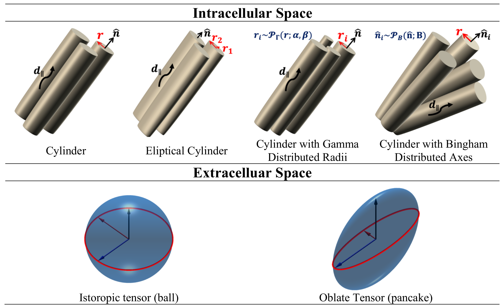
Figure 1. Intracellular and extracellular compartment models.
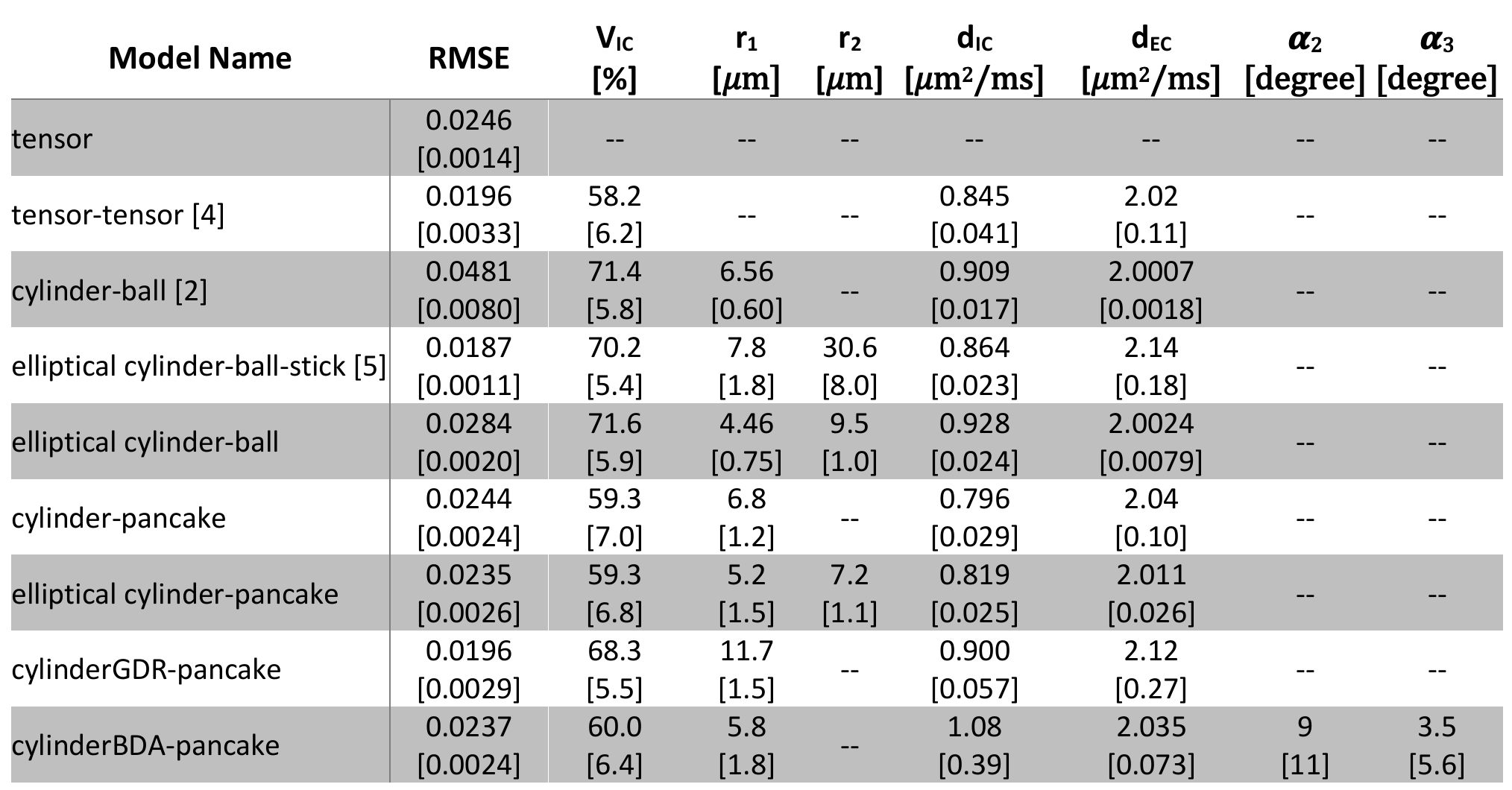
Table 1. Mean [STD] of Biophysical Parameters on a Slab from the Left Ventricle Wall. The 𝜶2 and 𝜶3 are the dispersion about the primary eigenvector in the sheetlet plane and sheetlet-normal plane. Reported physiological range from literature for intracellular, extracellular and vessels volume fractions are 65.0±3.6, 31.2±5.8, and 7.7±2.2 percent 8. Myocyte radii along the short and long axes are 6.0±0.7 and 15.3±1.0 𝜇m 7. Assuming a circular cross-section, the radius is 9.5±0.5 𝜇m 7.