Dominika Ciupek1, Maryam Afzali2, Fabian Bogusz1, Marco Pizzolato3,4, Derek K. Jones2, and Tomasz Pięciak1,5
1AGH University of Science and Technology, Kraków, Poland, 2Cardiff University Brain Research Imaging Centre (CUBRIC), School of Psychology, Cardiff University, Cardiff, United Kingdom, 3Department of Applied Mathematics and Computer Science, Technical University of Denmark, Kongens Lyngby, Denmark, 4Signal Processing Lab (LTS5), École polytechnique fédérale de Lausanne (EPFL), Lausanne, Switzerland, 5LPI, ETSI Telecomunicación, Universidad de Valladolid, Valladolid, Spain
1AGH University of Science and Technology, Kraków, Poland, 2Cardiff University Brain Research Imaging Centre (CUBRIC), School of Psychology, Cardiff University, Cardiff, United Kingdom, 3Department of Applied Mathematics and Computer Science, Technical University of Denmark, Kongens Lyngby, Denmark, 4Signal Processing Lab (LTS5), École polytechnique fédérale de Lausanne (EPFL), Lausanne, Switzerland, 5LPI, ETSI Telecomunicación, Universidad de Valladolid, Valladolid, Spain
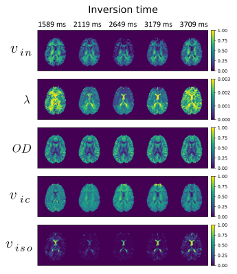
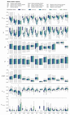
Most of the metrics of biophysical models are very sensitive to changes in TI values, therefore they can be used to analyse the brain microstructure only for high TIs.

Figure 1: Visual representation of SMT (intracellular volume fraction $$$v_{in}$$$ and intrinsic diffusivity $$$\lambda \ [\mathrm{mm}^2/\mathrm{s}]$$$) and NODDI (orientation dispersion index $$$OD$$$, intracellular volume fraction $$$v_{ic}$$$ and isotropic volume fraction $$$v_{iso}$$$) parameters under different inversion times.

Figure 2: Mean values and first and third quartiles for SMT (intracellular volume fraction $$$v_{in}$$$ and intrinsic diffusivity $$$\lambda \ [\mathrm{mm}^2/\mathrm{s}]$$$) and NODDI (mean orientation of Watson distribution $$$\mu = (\theta, \phi) \ [\mathrm{rad}]$$$, orientation dispersion index $$$OD$$$, intracellular volume fraction $$$v_{ic}$$$ and isotropic volume fraction $$$v_{iso}$$$) over regions-of-interests and changes in TI. WM - white matter, GM - grey matter, CSF - cerebrospinal fluid.