Zhixin Li1,2,3, Yue Wu1,2,3, Dongbiao Sun1,2,3, Jing An4, Qingle Kong5, Rong Xue1,2,3, Yan Zhuo1,2,3, and Zihao Zhang1,2,3
1State Key Laboratory of Brain and Cognitive Science, Institute of Biophysics, Chinese Academy of Sciences, Beijing, China, 2The Innovation Center of Excellence on Brain Science, Chinese Academy of Sciences, Beijing, China, 3University of Chinese Academy of Sciences, Beijing, China, 4Siemens Shenzhen Magnetic Resonance Ltd., Shenzhen, China, 5MR Collaboration, Siemens Healthcare Ltd, Beijing, China
1State Key Laboratory of Brain and Cognitive Science, Institute of Biophysics, Chinese Academy of Sciences, Beijing, China, 2The Innovation Center of Excellence on Brain Science, Chinese Academy of Sciences, Beijing, China, 3University of Chinese Academy of Sciences, Beijing, China, 4Siemens Shenzhen Magnetic Resonance Ltd., Shenzhen, China, 5MR Collaboration, Siemens Healthcare Ltd, Beijing, China
A
comprehensive framework was proposed to overcome the challenges in the automated
modeling of 3D cerebral small vessels with 7T TOF-MRA. The vasculature
and quantification of LSA were obtained in an automated
way, which facilitate related radiological studies in the future.
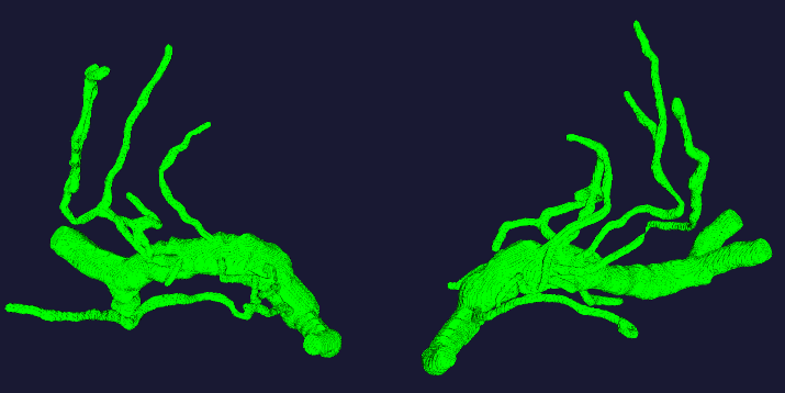
Figure 1. The 3D vasculature from 7T TOF-MRA obtained by our progressive method.
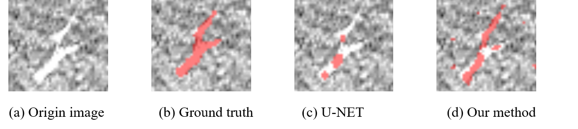
Figure 2. Comparison between the segmentation results of our method and U-NET. (a)
Origin image. (b) Ground truth. (c) Result of U-NET. (d) Result of our method.