Grant Hartung1, Joerg Pfannmoeller1, Avery J. L. Berman1, and Jonathan R. Polimeni1
1Athinoula A. Martinos Center for Biomedical Imaging, Boston, MA, United States
1Athinoula A. Martinos Center for Biomedical Imaging, Boston, MA, United States
We adapted realistic models of
cortical microvasculature to alter the topology of the vascular network and
performed dynamical simulations of blood flow and volume changes following
neural activity. We find that the topology changes cause qualitatively
different blood volume responses.
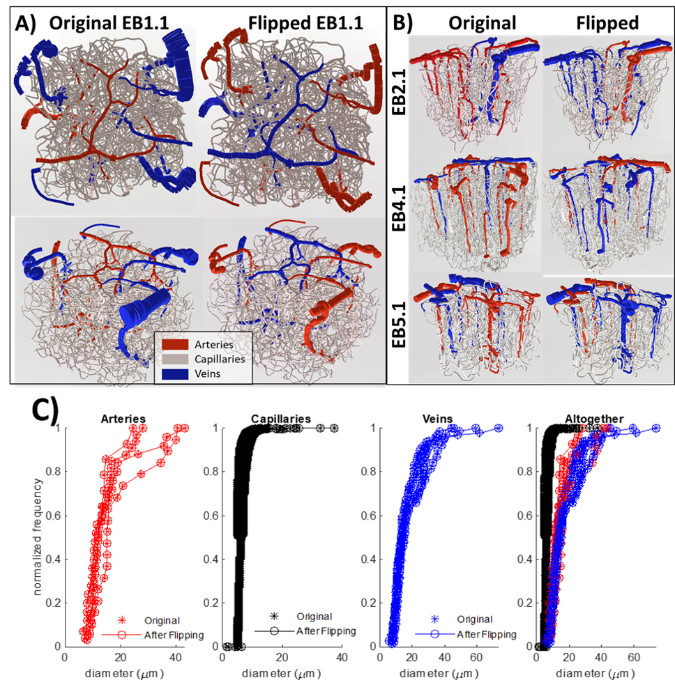
Figure
1. Visualization of A) the first network and B)
the other three networks both (Left Column) before and (Right Column) after the
anatomical swapping of arteries and veins. Not only was the labeling swapped
but the diameter spectra were also reassigned so that the newly assigned
arteries have the same diameter distribution as the previous arteries and
similarly the veins share the diameter spectra of the original veins. C) This
topological matching is also reflected by plotting the diameter spectra of the
arteries, capillaries, and veins from before and after swapping the anatomy.
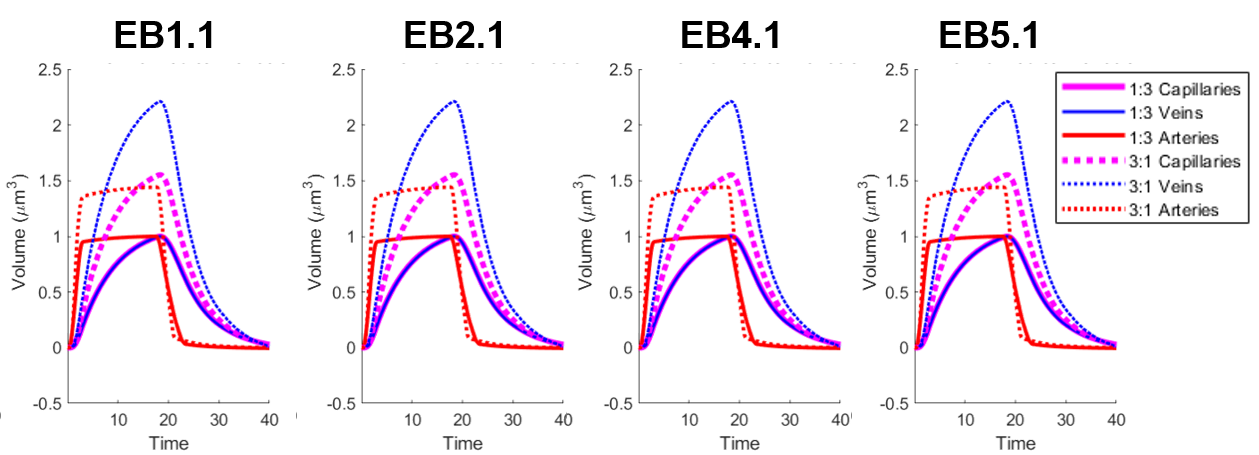
Figure
4. Visualization of the cerebral blood volume
(CBV) time course for all 4 VANs. The dilation in the experimental group (3:1
ratio) is significantly higher than the control counterpart in each case.