Martin John MacKinnon1,2,3,4, Yuncong Ma1,2,4, Sheng Song1,2,4, Tzu-Hao Harry Chao1,2,4, Tzu-Wen Winnie Wang1,2,4, SungHo Lee2,4, SungHo Lee1,2,4, Wei-Tang Chang2,5, and Yen-Yu Ian Shih1,2,4
1Center for Animal MRI, University of North Carolina at Chapel Hill, Chapel Hill, NC, United States, 2Biomedical Research Imaging Center, University of North Carolina at Chapel Hill, Chapel Hill, NC, United States, 3The Joint Department of Biomedical Engineering, University of North Carolina at Chapel Hill, Chapel Hill, NC, United States, 4Department of Neurology, University of North Carolina at Chapel Hill, Chapel Hill, NC, United States, 5Department of Radiology, University of North Carolina at Chapel Hill, Chapel Hill, NC, United States
1Center for Animal MRI, University of North Carolina at Chapel Hill, Chapel Hill, NC, United States, 2Biomedical Research Imaging Center, University of North Carolina at Chapel Hill, Chapel Hill, NC, United States, 3The Joint Department of Biomedical Engineering, University of North Carolina at Chapel Hill, Chapel Hill, NC, United States, 4Department of Neurology, University of North Carolina at Chapel Hill, Chapel Hill, NC, United States, 5Department of Radiology, University of North Carolina at Chapel Hill, Chapel Hill, NC, United States
We study the feasibility of using ZTE to detect functional activations
with endogenous contrast using a rat forepaw electrical stimulation
paradigm. We show that ZTE-fMRI has a 67% greater sensitivity than the
gold-standard BOLD-weighted EPI.
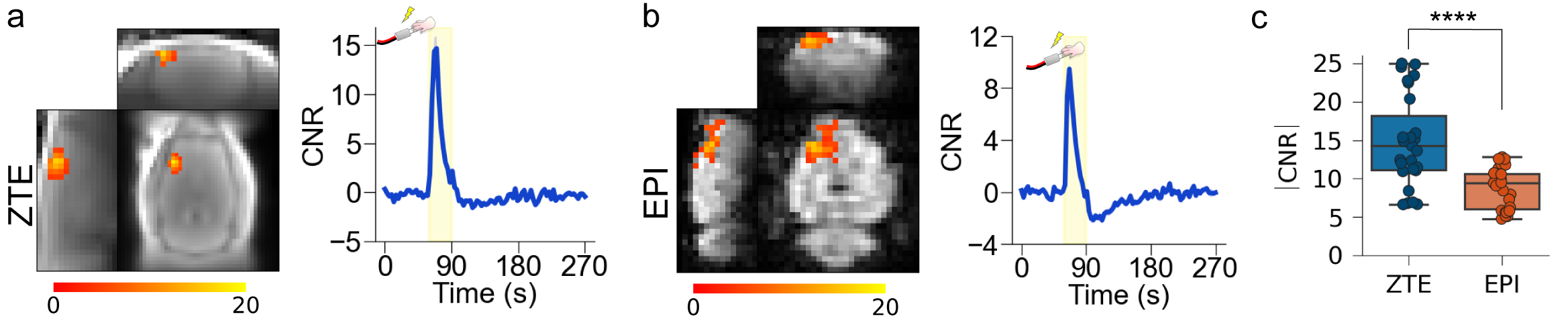
Figure 4.
ZTE and EPI functional
responses to forepaw electrical stimulation. a) ZTE-fMRI and b) BOLD-weighted
fMRI activation maps and averaged timcourses. Time courses were extracted from the mean of 3 voxel3 ROIs in contralateral S1. ZTE-fMRI exhibited a 67 % greater
CNR than that of BOLD-weighted EPI ( t(52)=4.80,P=1.37x10⁻⁵).
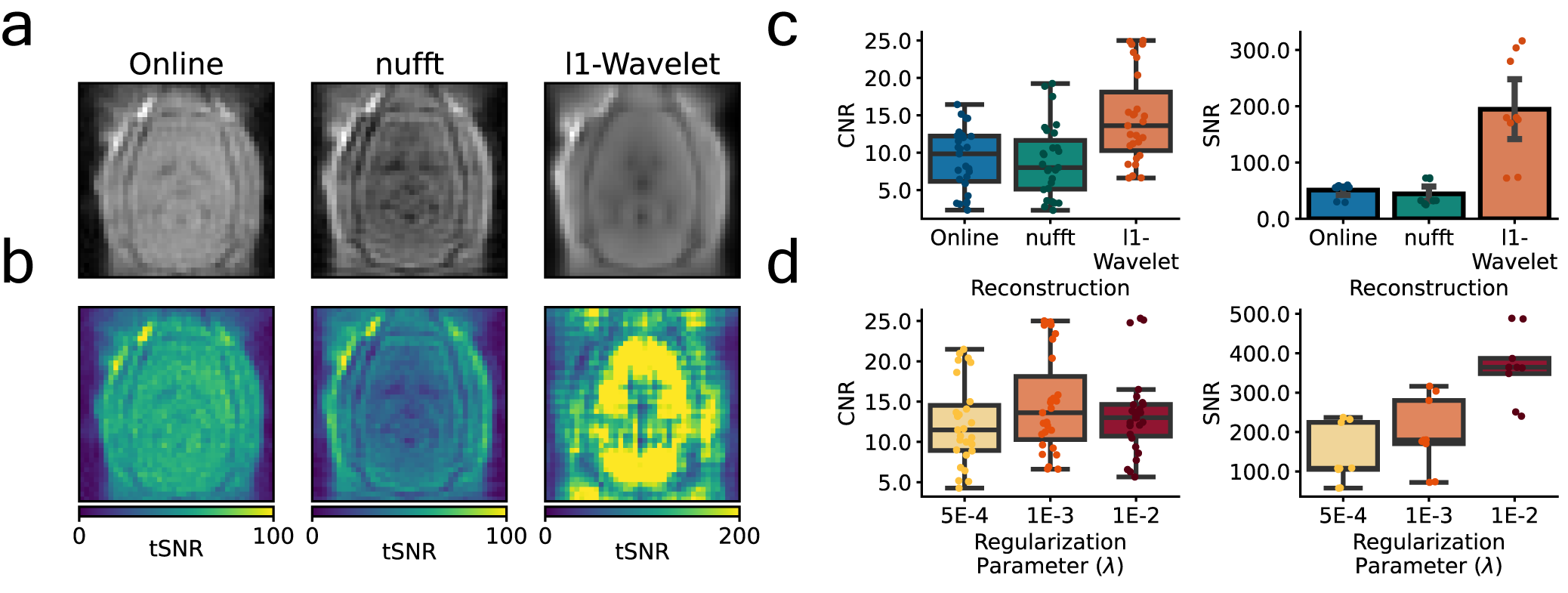
Figure 2: Effect of reconstruction algorithm on ZTE functional data. Coronal (scanner coordinate system) views of raw ZTE-fMRI data (a) following online reconstruction, nufft and l1-Wavelet regularization and (b) the corresponding tSNR maps. Comparison of CNR of evoked response and SNR of functional data during rat forepaw electrical stimulation (c). Effect of l1-wavelet regularization parameter on CNR and SNR of functional ZTE data. For both c) and (d) n=3 subjects, 27 trials.