Odélia Jacqueline Chitrit1, Qingjia Bao1, Silvia Chuartzman2, Noga Silkha2, Tali Kimchi2, and Lucio Frydman1
1Department of Chemical and Biological Physics, Weizmann institute of Science, Rehovot, Israel, 2Department of Neurobiology, Weizmann institute of Science, Rehovot, Israel
1Department of Chemical and Biological Physics, Weizmann institute of Science, Rehovot, Israel, 2Department of Neurobiology, Weizmann institute of Science, Rehovot, Israel
Spatiotemporal Encoding (SPEN) MRI was used in fully and non fully-refocused modes, to capture the activation of Olfactory Bulbs in mice, in response to odors. At the 15.2T field, the image quality largely exceeded that arising in GE or even SE EPI and responses on the order of 10% could be observed.
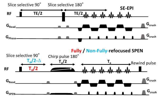
Figure 1: Representative
Spin Echo EPI (top) and Fully/Non-Fully-Refocused SPEN (bottom) acquisitions
used in this study. The latter included a 180˚ chirp pulse acting in the
presence of a gradient that encodes the more artifact-prone, low bandwidth
dimension, and lasts half the duration of the readout acquisition train Ta. This is preceded by a pre-encoding delay; if
set to Ta/2 full-refocusing is achieved, and T2*
effects are largely attenuated (red); if set to a shorter time (blue) a T2*
weighting is partially reintroduced.
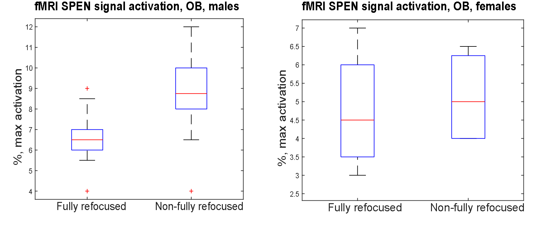
Figure 5: Box-whiskers plots describing the maximal signal activation
observed for the mice examined by SPEN in this work.