Shinho Cho1, Steen Moeller1, Mehmet Akçakaya2, Logan Dowdle1, Luca Vizioli1, Djaudat Idiyatullin1, Wei Chen1, and Kâmil Uğurbil1
1Center for Magnetic Resonance Research and Department of Radiology, University of Minnesota, Minneapolis, MN, United States, 2Center for Magnetic Resonance Research and Department of Electrical and Computer Engineering, University of Minnesota, Minneapolis, MN, United States
1Center for Magnetic Resonance Research and Department of Radiology, University of Minnesota, Minneapolis, MN, United States, 2Center for Magnetic Resonance Research and Department of Electrical and Computer Engineering, University of Minnesota, Minneapolis, MN, United States
NORDIC denoising to suppress thermal noise in zero-mean
Gaussian distribution, yielding improved functional CNR and T-statistics with
minimal increase in spatial smoothing and equivalent results in the signal
stability of 3-4 times averaging of single fMRI acquisition.
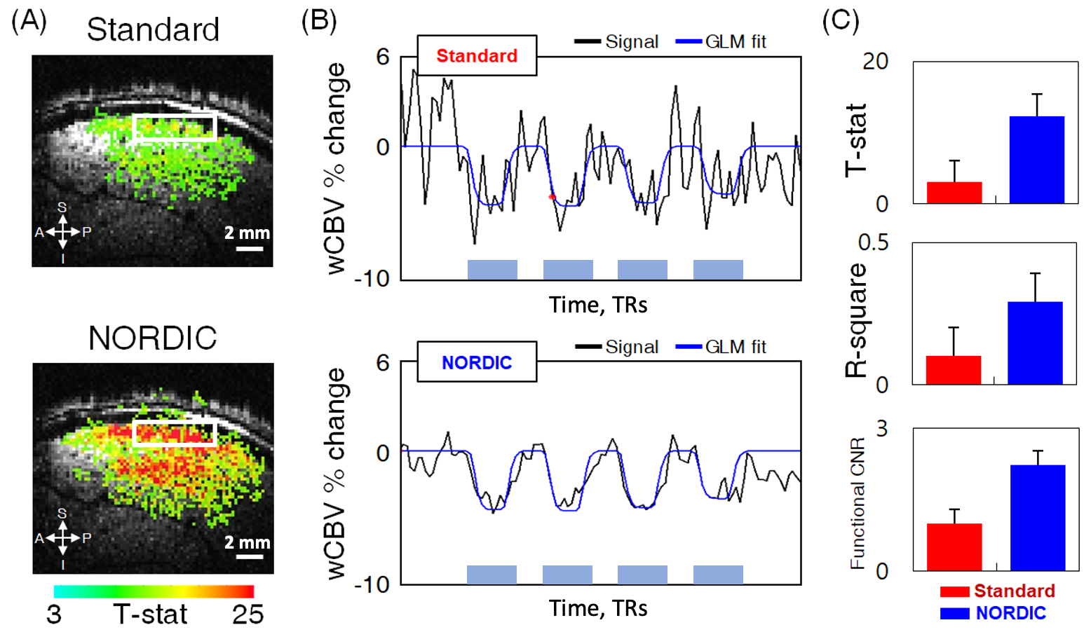
Fig.
1. wCBV fMRI results improved by NORDIC denoising. (A) wCBV activation map (T-statistics (T ≥ 3), sagittal slice, single subject) induced by stimuli.
(B) The mean time course from the region-of-interest (ROI) denoted by white
rectangles in (A); stimulus-induced wCBV signal changes shown during
stimulus on epochs (blue/gray rectangles). The raw EPI signal times series
(black) is shown together with the GLM fit (blue). (C) Effect of NORDIC on
the average T-stats in the
region-of-interest (ROI), R-square, and functional CNR; the error
bars are SEM.
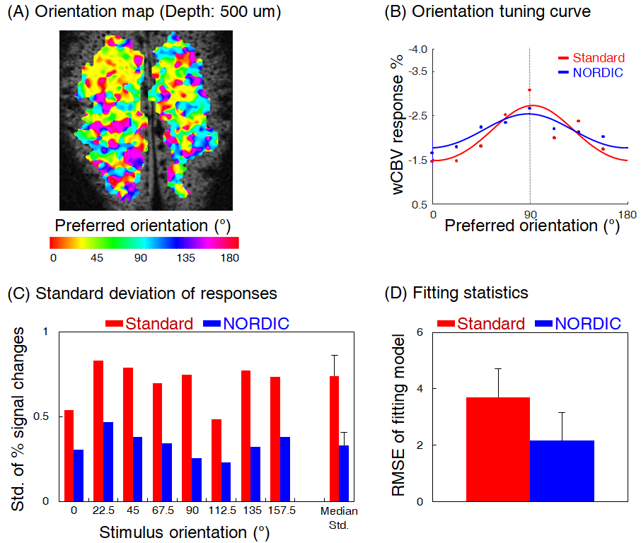
Fig. 4. Orientation preference
mapping and statistics improved before and after NORDIC denoising. (A) Orientation preference mapped by NORDIC denoised wCBV responses to
8-orientation (single subject); colors indicate the orientation
preference in degree. (B) Representative tuning curve of a single voxel before-
(red) and after-NORDIC denoising (blue). (C-D)
The standard deviations of multiple wCBV responses repeatedly measured and goodness-of-fit of curve fitting (root-mean-square-error) were compared between before and after NORDIC denoising.