Nmachi Anumba1,2, Wenju Pan1,2, Eric Maltbie1,2, and Shella Keilholz1,2
1Biomedical Engineering, Emory University, Atlanta, GA, United States, 2Biomedical Engineering, Georgia Institute of Technology, Atlanta, GA, United States
1Biomedical Engineering, Emory University, Atlanta, GA, United States, 2Biomedical Engineering, Georgia Institute of Technology, Atlanta, GA, United States
We performed a voxel-wise analysis of the BOLD global signal in rat rs-fMRI and found that contributions to the global signal are not uniformly distributed throughout the brain. We also compared the signal to nonneural tissue and found that it contains attributes that are unique to the brain.
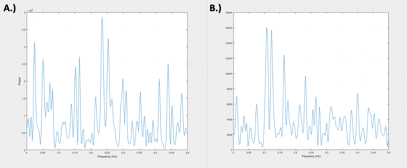
Figure 1. Averaged Global Signals for Muscle and Brain. A.) Shows the frequency power spectrum of the averaged brain global signal of 49 scans, each 10 minutes in length, taken across 8 rats. B.) Shows the frequency power spectrum of the averaged muscle “global signal” of 49 scans, each 10 minutes in length, taken across 8 rats. It is important to note the relative difference in magnitude between the two signals with the muscle global signal displaying much lower power.
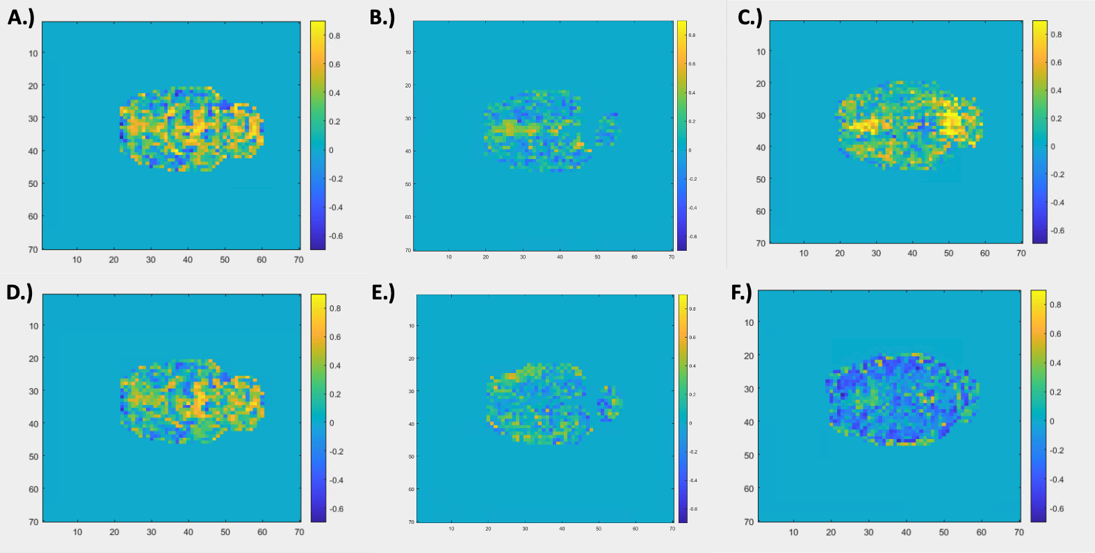
Figure 2. Voxel-wise Correlation to Brain and Muscle Global Signal. Panels A – C show a voxel-wise map of Pearson correlation coefficients for 3 individual rats for which the timecourse of each voxel was compared to the global signal of the brain. Panels D – E show the same analysis comparing voxels to the “global signal” of the muscle. Panels D – E display the same rat and slice number as the corresponding panel in the first row (A – C, respectively). Correlation values for muscle to brain global signals for each rat, from left to right, were R = 0.7313, R = -0.0839, and R = -0.1345, respectively.