Naman Jain1, Atena Akbari1, Markus Barth1,2, and Kai-Hsiang Chuang1,3
1Centre for Advanced Imaging, The University of Queensland, Brisbane, Australia, 2School of Information Technology and Electrical Engineering, The University of Queensland, Brisbane, Australia, 3Queensland Brain Institute, The University of Queensland, Brisbane, Australia
1Centre for Advanced Imaging, The University of Queensland, Brisbane, Australia, 2School of Information Technology and Electrical Engineering, The University of Queensland, Brisbane, Australia, 3Queensland Brain Institute, The University of Queensland, Brisbane, Australia
We tested the feasibility of CBV - weighted VASO fMRI in mouse models and results sho that it is possible to do VASO in mouse. An expected negative signal change was observed as reported in the literature.
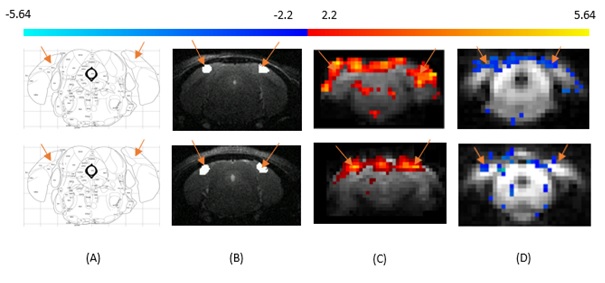
Figure
1: Primary visual cortex is specified with reference to
Paxinos and Franklin Mouse Brain Atlas (Figure 1A), V1 is marked with orange
arrows in T2-weighted structural scan (Figure 1B). Primary visual cortex
indicated by the arrows. (C) BOLD and (D) VASO functional maps of mouse primary
visual cortex. Row 1 (Top) and Row 2 (Bottom) are the results acquiried from
two different mice.
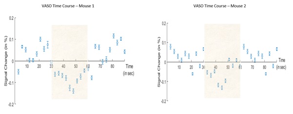
Figure 2: VASO signal time course of averaged across all
stimulation blocks (shaded area) of 4 runs for 2 different mice, errorbars
represents the standard error of mean.