Bruno Pradier1,2, Lydia Wachsmuth1, Daniel Segelcke2, Nina Nagelmann1, Esther Pogatzki-Zahn2, and Cornelius Faber1
1Department of Clinical Radiology, University Hospital Münster, Münster, Germany, 2Department of Anesthesiology, University Hospital Münster, Münster, Germany
1Department of Clinical Radiology, University Hospital Münster, Münster, Germany, 2Department of Anesthesiology, University Hospital Münster, Münster, Germany
We find that brain states quickly reach a
steady state after switching anesthetic regimen. We show that most changes in
functional connectivity were relative to the initial isoflurane anesthesia. We
conclude that brain states and networks are stable from 30min after switching
anesthetics.
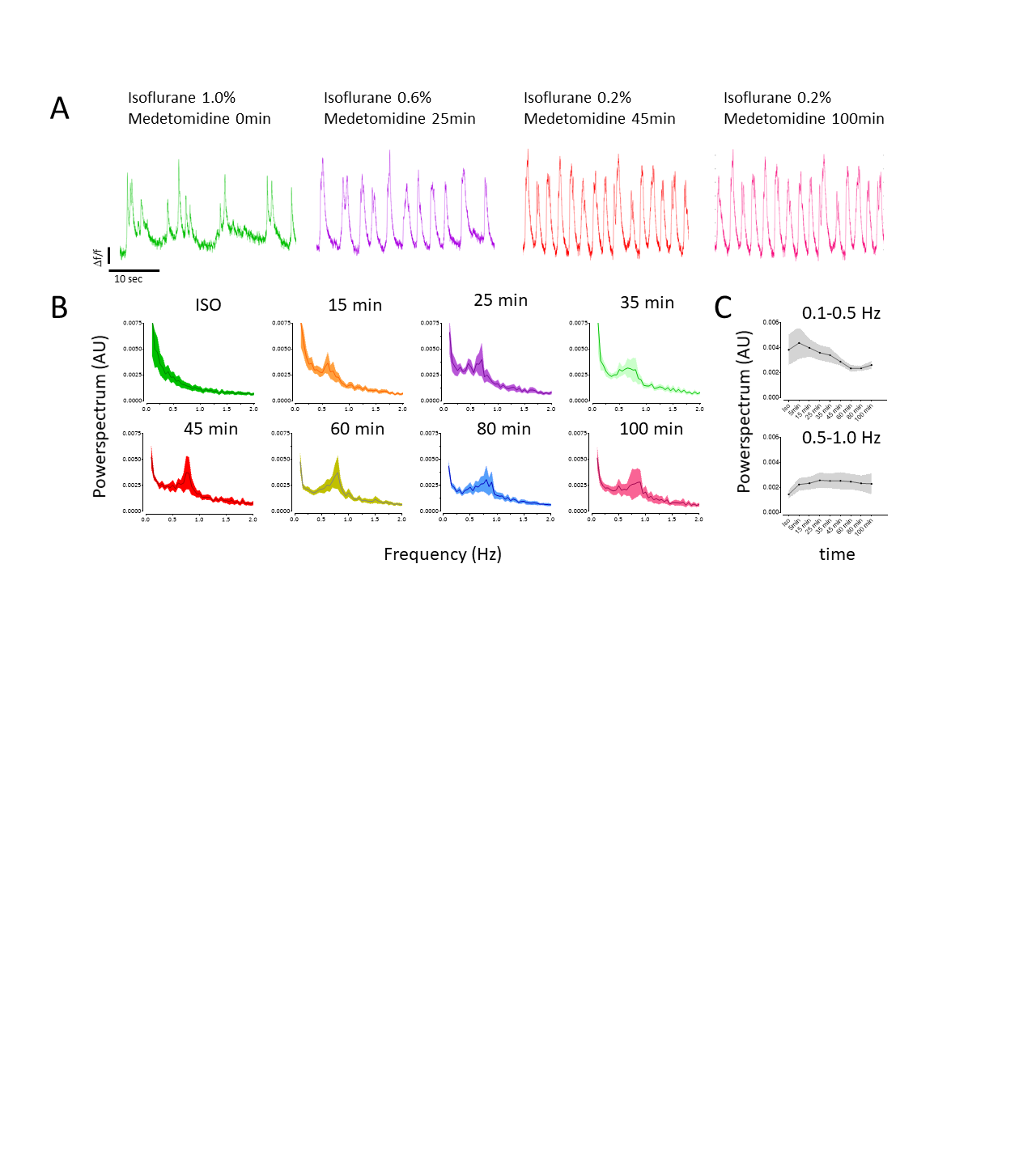
Calcium recordings show different brain
states in S1HL depending on anesthetic condition. (A) Under ISO anesthesia,
calcium transients show frequent UP-DOWN transitions (green) while changing to
a persistent state at 25 (purple), 45 (red), and 100 (pink) minutes after
switching to ISO/MED anesthesia. (B) Fourier-transformed calcium transients
show an emerging peak between 0.5Hz and 1.0 Hz 15 to 100 minutes after
switching anesthetic regimens. (C) Frequencies of calcium transients between
0.1-0.5Hz decreased (p=0.002), while frequencies from 0.5-1.0Hz increased (p=0.02).
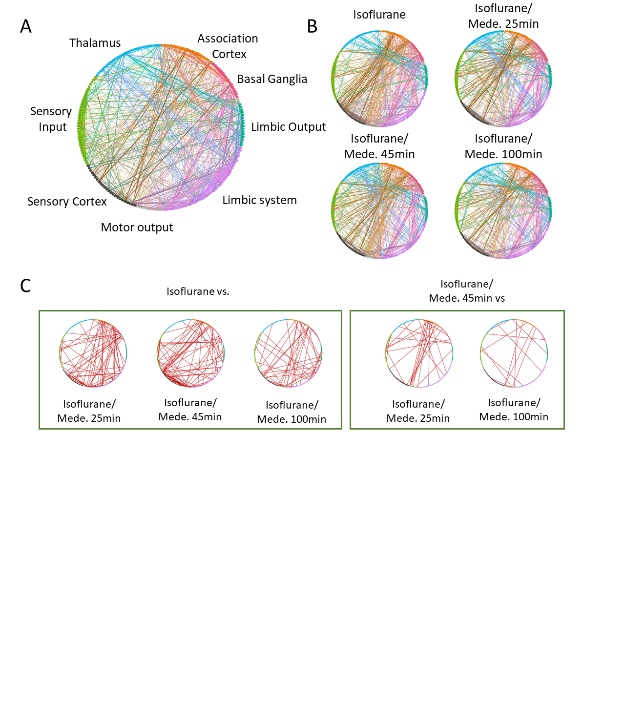
Changes in functional connectivity were
mostly related to a decrease in ISO concentration. (A) Circular network
representation. Each dot represents a brain region, color codes functional
groups, and lines represent functional connectivity based on pearson’s
correlation coefficient. (B) Averaged networks for each group. (C) Differences
in networks, obtained from statistical analysis of ISO vs. later time points
(left panel) and ISO/MED 45 min vs. ISO/MED 25min and ISO/MED 100 min (right panel).