Tomokazu Tsurugizawa1, Boucif Djemai2, and Kazumi Kasahara1
1Human Informatics and Interaction Research Institute, National Institute of Advanced Industrial Science and Technology (AIST), Tsukuba, Japan, 2NeuroSpin/CEA-Saclay, Gif-sur-Yvette, France
1Human Informatics and Interaction Research Institute, National Institute of Advanced Industrial Science and Technology (AIST), Tsukuba, Japan, 2NeuroSpin/CEA-Saclay, Gif-sur-Yvette, France
The peak of positive BOLD responses in the
contralateral S1 was correlated with the peak of negative BOLD response in the bilateral
barrel cortices during the somatosensory stimulation in mice.
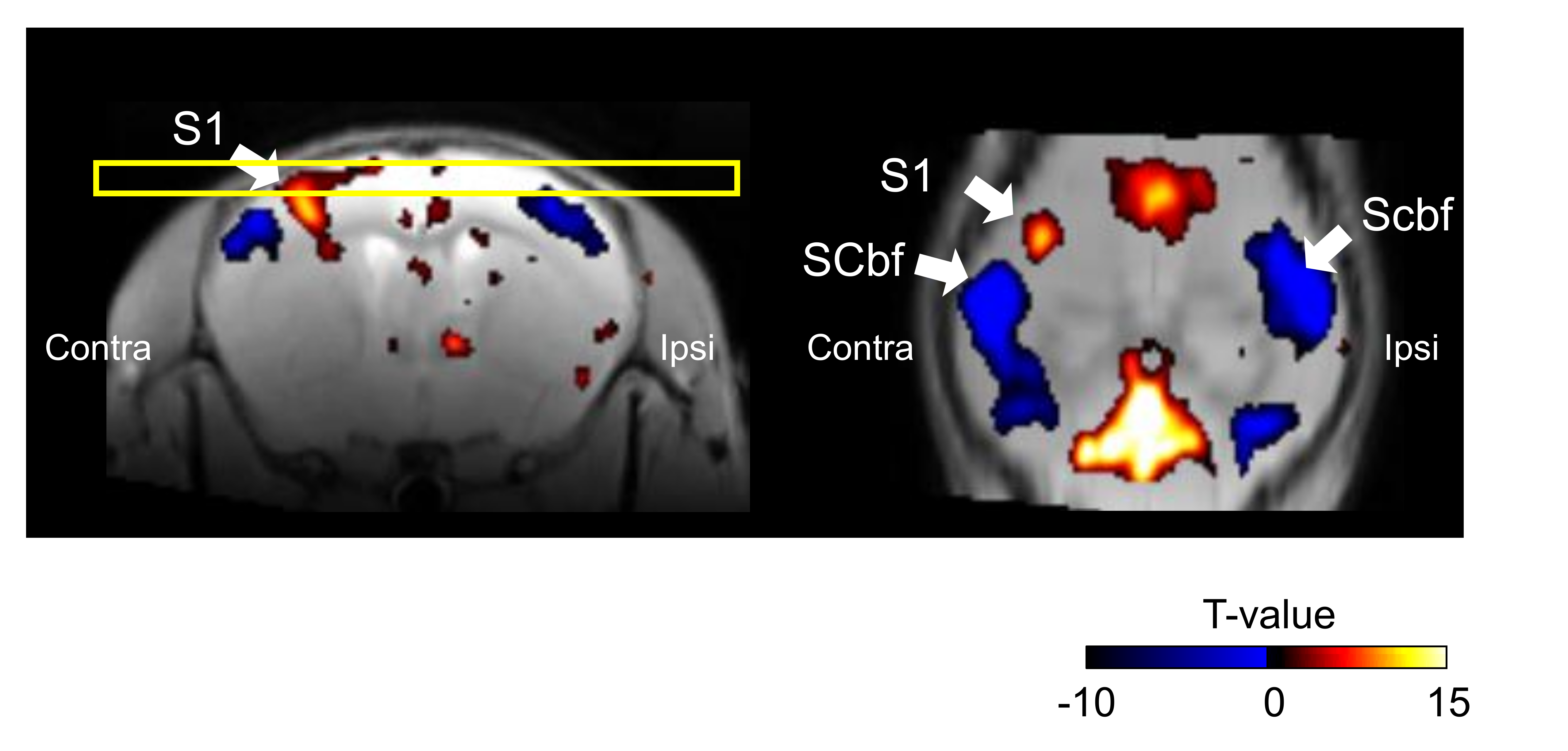
Figure
1
Group analysis of BOLD signal changes by the somatosensory stimulation
in mice (n=7, p<0.05, FDR-corrected). Yellow rectangle shows the horizontal
plane in right panel. Color bar indicates the t-values. SCbf, somatosensory
cortex of barrel field; S1; primary somatosensory cortex.
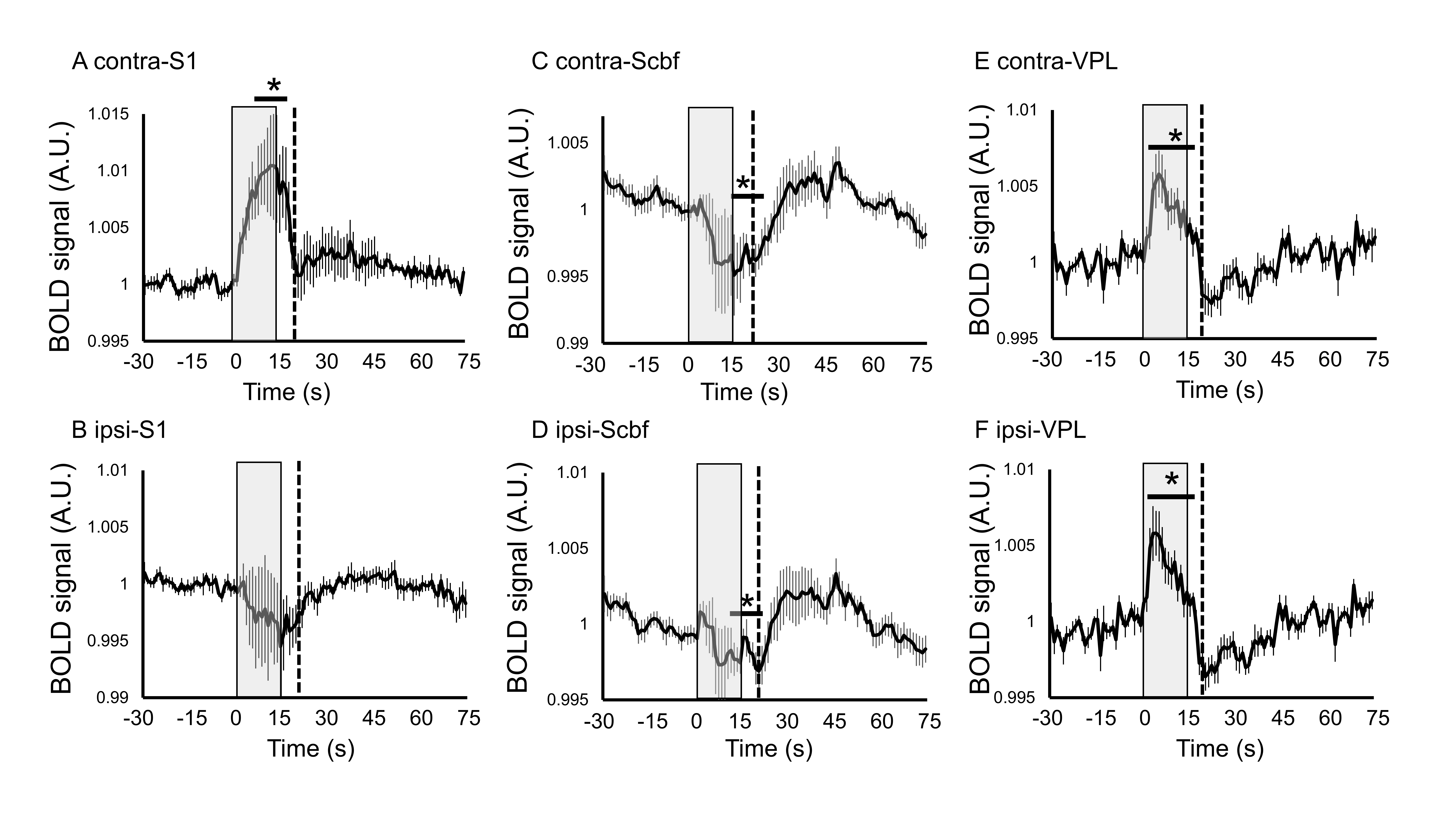
Figure
2 Averaged time-course of
BOLD signal changes in (A) contralateral primary somatosensory cortex (contra-S1),
(B) ipsilateral primary somatosensory cortex (ipsi-S1), (C) contralateral
primary somatosensory cortex barrel field (contra-Scbf), (D) ipsilateral
primary somatosensory cortex barrel field (ipsi-Scbf). (E) contralateral ventral
posterolateral nucleus (contra-VPL), (F) ipsilateral ventral posterolateral
nucleus (ipsi-VPL). Dotted line shows 5 seconds after the end of stimulation. Data
are mean ±
SEM. *p < 0.05, compared to averaged baseline.