Mohit H Adhikari1, Tamara Vasilkovska1, Dorian Pustina2, Longbin Liu2, Roger Cachope2, Haiying Tang2, Celia Dominguez2, Ignacio Munoz-Sanjuan2, Annemie Van der Linden1, and Marleen Verhoye1
1Biomedical Sciences, University of Antwerp, Antwerp, Belgium, 2CHDI Foundation, Princeton, NJ, United States
1Biomedical Sciences, University of Antwerp, Antwerp, Belgium, 2CHDI Foundation, Princeton, NJ, United States
Transient brain states during brain's spontaneous state measured by fMRI are altered in the early & manifest stages of Huntington's disease (HD) in mice. Their spatial properties accurately predict the identity of diseased mice making them promising candidates for a fMRI biomarker of HD.
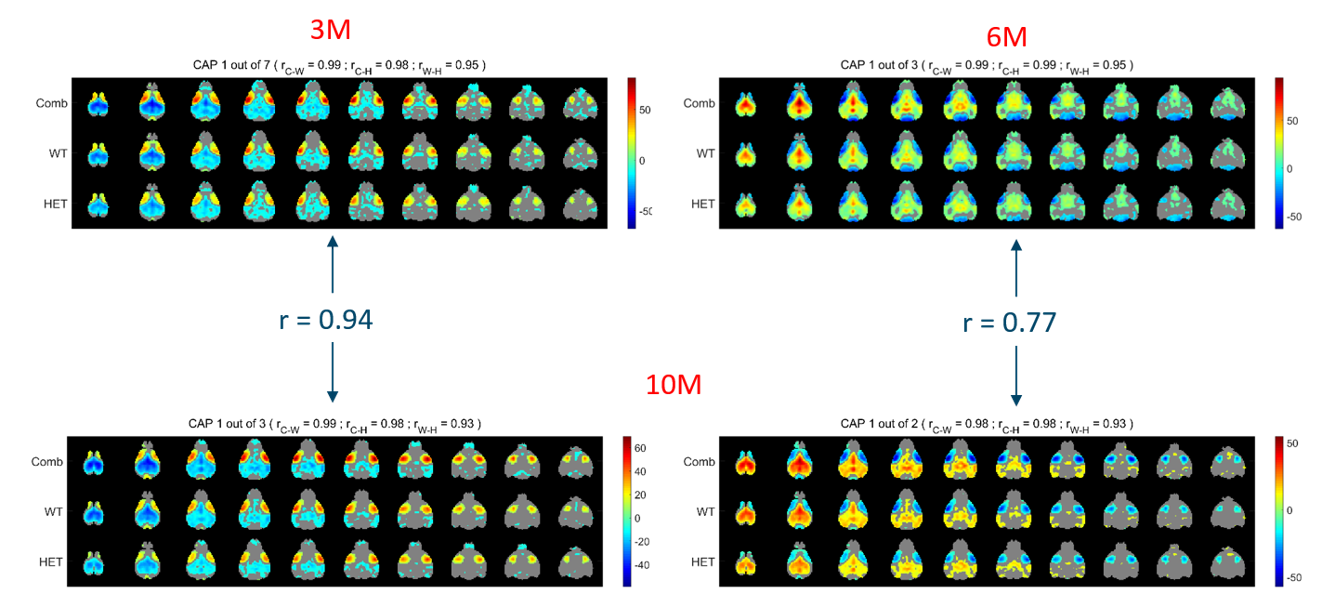
One-sample T-test statistic maps, derived from its occurrences in the combined (top), WT (middle), and HET (bottom) image-series, for two prototype CAPs with significantly reduced duration and/or occurrence in the HET group at the 10-month time point. At the 3 and 6-month time points, only one of these two patterns showed significantly reduced occurrence and duration in the HET group respectively. Left panel CAPs show simultaneous co-deactivation & co-activation of default mode-like and latero-cortical networks in mice while right panels show the reverse pattern.
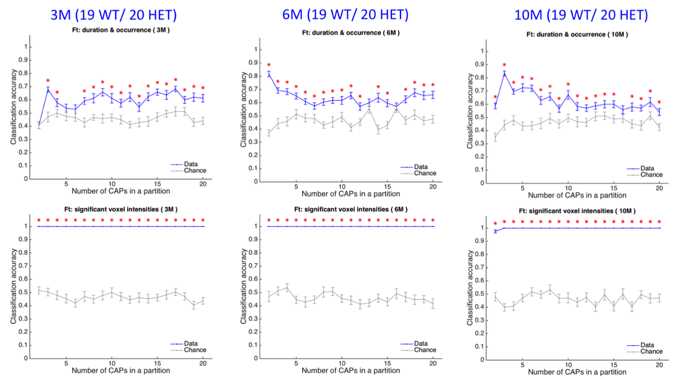
Classification accuracy (blue, mean +/- SEM) using duration and occurrence (top panels), and BOLD signals of voxels with significant activations (bottom panels) of each CAP within a partition as a function of partitions of the combined image series at the 3-month time point (left panels), 6-month time point (middle panels) & at the 10-month time point (right panels). The grey curve shows the corresponding chance-level accuracy (mean +/- SEM) and the red asterisk indicates significantly higher mean accuracy than the chance level, after correcting for multiple comparisons.