Teng Ma1,2,3, Xunda Wang1,2, Eddie C. Wong1,2, Pit Shan Chong4, Sanchal Sanchayyan4, Lee Wei Lim4, Pek-Lan Khong3, Ed X. Wu1,2, and Alex T. L. Leong1,2
1Laboratory of Biomedical Imaging and Signal Processing, The University of Hong Kong, Hong Kong SAR, China, 2Department of Electrical and Electronic Engineering, The University of Hong Kong, Hong Kong SAR, China, 3Department of Diagnostic Radiology, Li Ka Shing Faculty of Medicine, The University of Hong Kong, Hong Kong SAR, China, 4School of Biomedical Sciences, Li Ka Shing Faculty of Medicine, The University of Hong Kong, Hong Kong SAR, China
1Laboratory of Biomedical Imaging and Signal Processing, The University of Hong Kong, Hong Kong SAR, China, 2Department of Electrical and Electronic Engineering, The University of Hong Kong, Hong Kong SAR, China, 3Department of Diagnostic Radiology, Li Ka Shing Faculty of Medicine, The University of Hong Kong, Hong Kong SAR, China, 4School of Biomedical Sciences, Li Ka Shing Faculty of Medicine, The University of Hong Kong, Hong Kong SAR, China
By optogenetically stimulating the excitatory projection neurons in olfactory
bulb (OB), we revealed that OB neural activity propagated
to regions that are associated with higher-order cognition, reward-related
behaviors and multisensory processing.
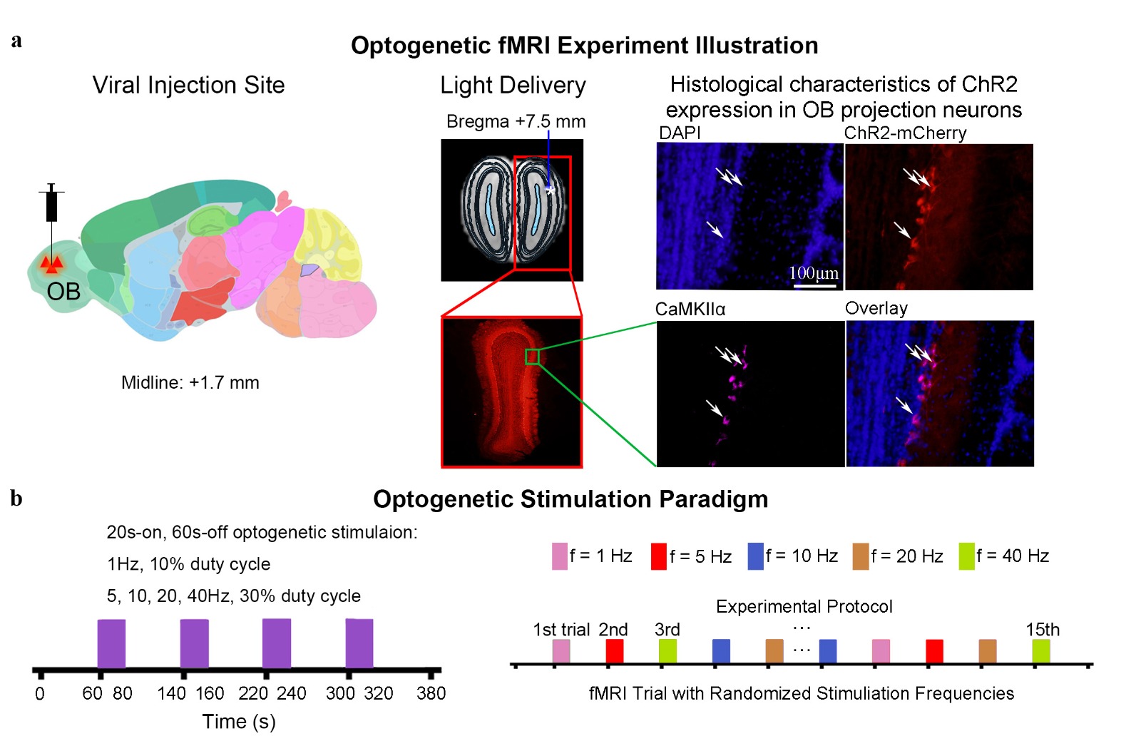
Figure
1. Experimental setup for optogenetic stimulation and histological characterization
of viral expression in OB excitatory neurons.
(a) Illustration of the location of viral injection (left) the fiber
implantation in (middle) T2-weighted anatomical image and the CamKIIα::ChR2
expression in excitatory neurons of OB. (b) Schematic illustrating the
optogenetic stimulation paradigm: consisting of four 20s-on and 60s-off blocks
(1Hz, 10% duty cycle; 5, 10, 20, 40Hz, 30% duty cycle; 40mW/mm2).
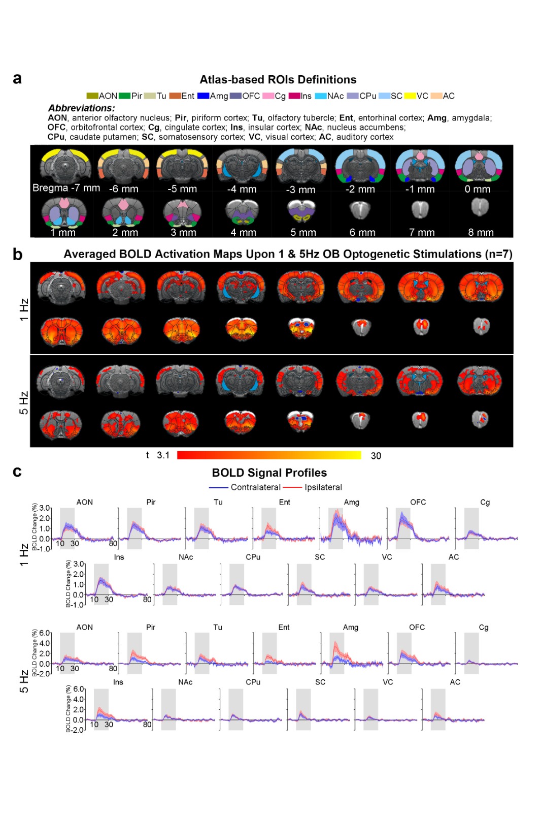
Figure
2. Robust brain-wide activations upon low frequency optogenetic stimulation of
OB projection neurons. (a) Regions-of-interests
(ROIs) definition based on atlas. (b) Averaged activations maps of
optogenetic stimulation in OB at 1Hz and 5Hz (n=7;
t>3.1, corresponding to p<0.001). (c) The respective BOLD signal
profiles extracted from ROIs at 1Hz and 5Hz. Error bars indicate ± SEM.