Eddie C. Wong1,2, Teng Ma1,2,3, Xunda Wang1,2, Pek-Lan Khong3, Ed X. Wu1,2, and Alex T.L. Leong1,2
1Laboratory of Biomedical Imaging and Signal Processing, The University of Hong Kong, Hong Kong SAR, China, 2Department of Electrical and Electronic Engineering, The University of Hong Kong, Hong Kong SAR, China, 3Department of Diagnostic Radiology, Li Ka Shing Faculty of Medicine, The University of Hong Kong, Hong Kong SAR, China
1Laboratory of Biomedical Imaging and Signal Processing, The University of Hong Kong, Hong Kong SAR, China, 2Department of Electrical and Electronic Engineering, The University of Hong Kong, Hong Kong SAR, China, 3Department of Diagnostic Radiology, Li Ka Shing Faculty of Medicine, The University of Hong Kong, Hong Kong SAR, China
We combined fMRI and optogenetic
stimulation of vestibular excitatory neurons to visualize two distinct
brain-wide functional organization of central vestibular pathways originating
from two major vestibular subnuclei, the superior vestibular nucleus and medial vestibular nucleus.
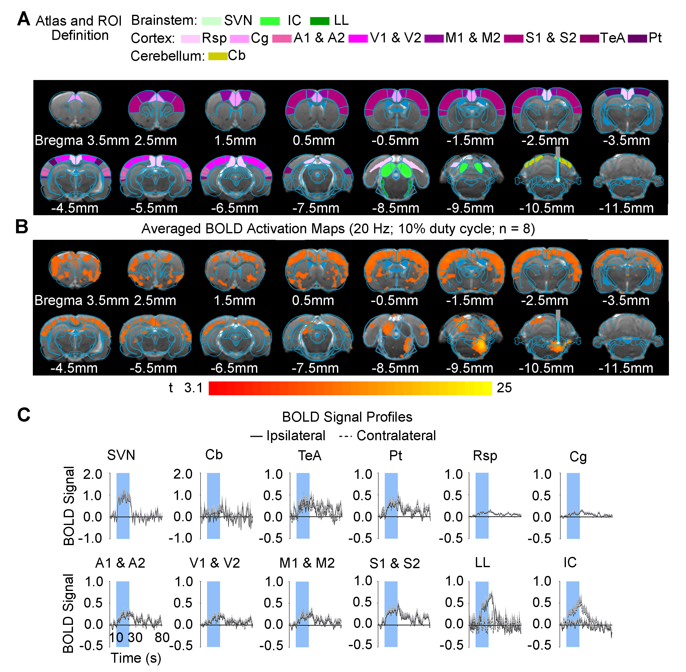
Figure
2. Brain-wide activation at cortical and
subcortical regions upon optogenetic stimulation of SVN excitatory neurons. (A)
Illustration of atlas-based region of interest (ROI) definitions in the
vestibular, midbrain, cerebellum, and cortical regions. (B) Averaged
BOLD activation maps at 20-Hz optogenetic stimulation at SVN (n = 8; P <
0.001; asterisk indicates the stimulation site). (C) BOLD signal
profiles extracted from ROIs defined in A (error bars indicate ± SEM).
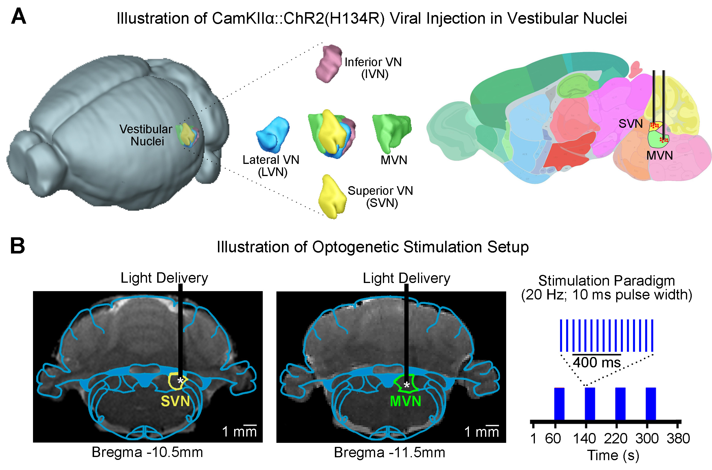
Figure 1.
(A) 3D illustration of the four vestibular subnuclei (top-left)
and illustration of CfR2-transfected MVN and SVN excitatory neurons (top-right).
(B) Illustration of the
stimulation site in ChR2 SVN (bottom-left) and ChR2 MVN (bottom-middle)
neurons during optogenetic fMRI experiments (bottom-right). T2-weighted
anatomical MRI image showing the location of the implanted optical fiber
(asterisk, stimulation site; right).