Hyeon-Joong Kim1, Ryan S Clement2, Roger B Bagwell2, Tirko N Natasha2, Yen-Yu Ian Shih1, and Sung-Ho Lee1
1Center for Animal MRI, Biomedical Research Imaging Center, University of North Carolina, Chapel Hill, NC, United States, 2Actuated medical, Bellefonte, PA, United States
1Center for Animal MRI, Biomedical Research Imaging Center, University of North Carolina, Chapel Hill, NC, United States, 2Actuated medical, Bellefonte, PA, United States
Stimulating NAc shell increased the BOLD responses in medial prefrontal cortex (mPFC), anterior cingulate cortex, and amygdala while the stimulating NAc core increased mostly mPFC. These are elucidated by using Deep Brain Stimulation-Functional Magnetic Resonance Imaging.
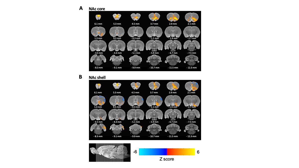
Figure 2. BOLD responses to NAc-DBS occurred separately between the NAc core and shell. Averaging 9 animals with NAc core stimulation (A) and NAc shell stimulation (B) respectively. All subjects were scanned with 200 μA DBS. (p < 0.05, voxel size > 40)
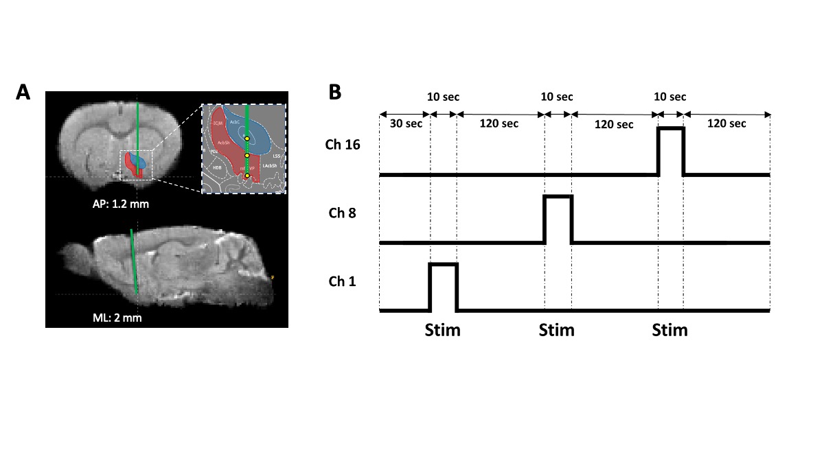
Figure 1. The Positioning Validation of electrode implantation and a stimulation paradigm. (A) Representative image from one subject for electrode positioning. (n = 9) (B) Stimulation paradigm. A single stimulation was applied to each channel during a single scan session. After the first 30 seconds of rest, stimulation was given 3 times for 10 seconds each, and 120 seconds of rest was given between stimulations. These 3 stimuli were given to channels 1, 8, and 16 respectively, and repeated 5 times per one animal.