Edward Peake1,2,3, Stefan Pszczolkowski1,2,3, Christoph Arthofer2,3,4, and Dorothee P Auer1,2,3
1NIHR Nottingham Biomedical Research Centre, University of Nottingham, Nottingham, United Kingdom, 2Radiological Sciences, Division of Clinical Neuroscience, University of Nottingham, Nottingham, United Kingdom, 3Sir Peter Mansfield Imaging Centre, University of Nottingham, Nottingham, United Kingdom, 4Nuffield Department of Clinical Neuroscience, University of Oxford, Oxford, United Kingdom
1NIHR Nottingham Biomedical Research Centre, University of Nottingham, Nottingham, United Kingdom, 2Radiological Sciences, Division of Clinical Neuroscience, University of Nottingham, Nottingham, United Kingdom, 3Sir Peter Mansfield Imaging Centre, University of Nottingham, Nottingham, United Kingdom, 4Nuffield Department of Clinical Neuroscience, University of Oxford, Oxford, United Kingdom
MRI improves prediction of knee replacement surgery. A radiomic signature derived from MRI features had a cox hazard ratio of 7.5 (p = 2e-28, 95% CI 7.1 – 7.9) which was higher than established clinico-demographic multivariate prediction model with cox hazard ratio 5.9 (p = 2e-28, 95%CI 5.6 – 6.2).
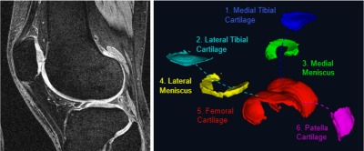
Left | DESS knee MRI with a 140mm field of view; 0.7mm slice thickness and matrix of 384×384 with 160 slices and an acquisition time of 10.6 mins. Right | Cartilage components segmented using our fully automated segmentation method based on U-convolutional neural networks.
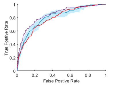
ROC curves for prediction of total knee replacement within 5-years. Blue | Radiomic signature with a C-index of 0.81 (95% CI, 0.76 – 0.84). Purple | Cox risk model including clinico-demographic covariates and the radiomic signature with a C-index of 0.85 (95% CI, 0.82 – 0.87). Red | cox risk model with clinico-demographic features alone had a C-index of 0.79 (95% CI, 0.75 – 0.83).