Seyed Amir Mirmojarabian1, Victor Casula1, Olli-Pekka Aro1, Henning Henschel2, Miika T Nieminen1,3,4, and Timo Liimatainen1,3
1Research Unit of Medical Imaging, Physics and Technology, University of Oulu, Oulu, Finland, 2Department of Medicinal Chemistry, Uppsala University, uppsala, Sweden, 3Medical Research Center, Oulu University Hospital, Oulu, Finland, 4Department of Diagnostic Radiology,, Oulu University Hospital, Oulu, Finland
1Research Unit of Medical Imaging, Physics and Technology, University of Oulu, Oulu, Finland, 2Department of Medicinal Chemistry, Uppsala University, uppsala, Sweden, 3Medical Research Center, Oulu University Hospital, Oulu, Finland, 4Department of Diagnostic Radiology,, Oulu University Hospital, Oulu, Finland
Relaxation along fictitious field with pulse duration close to 2.8 ms efficiently
capture lack of -OH group in degraded compared to intact cartilage.

Figure 2: T2 map and TRAFF
relaxation time maps with implemented pulse durations (PD) from two cartilage
specimen; red rectangle shows areas of degraded cartilage and yellow rectangle
points out to intact cartilage in T2 map.
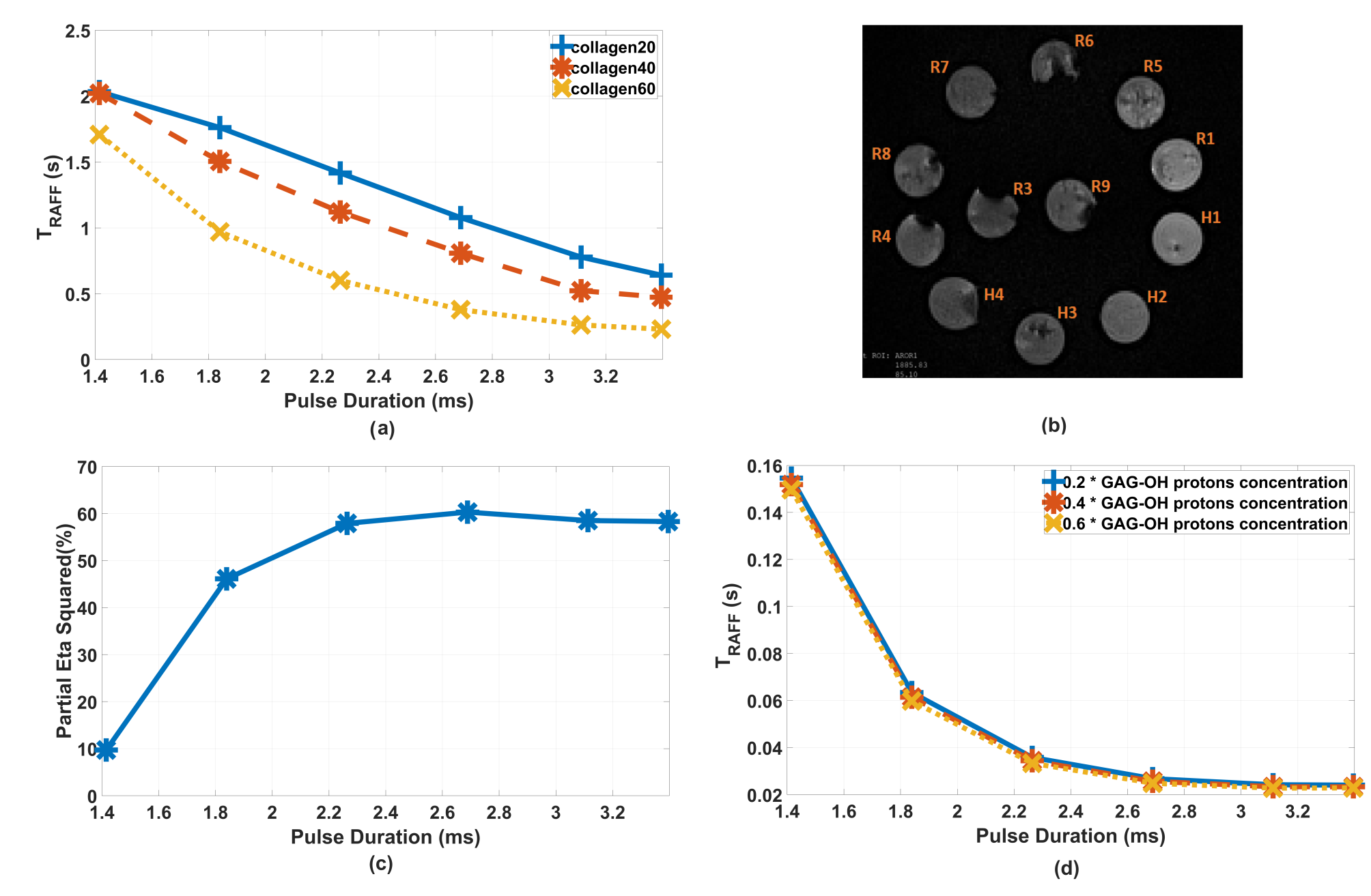
Figure 1: a) TRAFF
of collagen phantom measurements. b) T2 image of collagen phantoms. c) Partial
eta square percentage, representing TRAFF relaxation time
contrast/variation accounted by collagen concentration. d) Bloch-McConnell
simulated TRAFF.