Jiajia Li1, Shufang Wei1, Xianchang Zhang2, Jing An3, and Yinghui Ge1
1Fuwai Central China Cardiovascular Hospital, Zhengzhou, China, 2MR Collaboration, Siemens Healthcare Ltd., Beijing, China, Beijing, China, 3Siemens Shenzhen Magnetic Resonance Ltd, Shenzhen, China
1Fuwai Central China Cardiovascular Hospital, Zhengzhou, China, 2MR Collaboration, Siemens Healthcare Ltd., Beijing, China, Beijing, China, 3Siemens Shenzhen Magnetic Resonance Ltd, Shenzhen, China
T2*
mapping can detect early iron deposition caused by repeated bleeding in
patients with hemophilia and could be used as a sensitive biomarker to detect
early cartilage changes and develop preventative treatment plans.
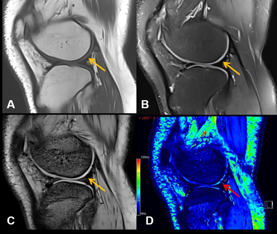
Images from one representative participant (male, 17 years old) with
hemophilic arthropathy. Conventional (A) T1WI. (B) PDWI. (C) T2*-anatomical
image show relatively complete articular surface (yellow arrow) without damage.
(D) T2* mapping shows uneven distribution in the cartilage and the decreases
(red arrow) of cartilage T2* value indicate damage.
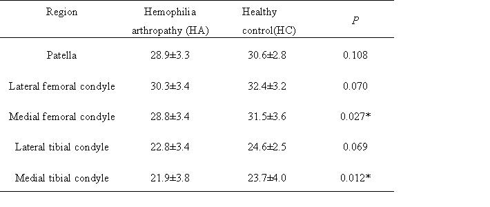
T2* values of articular cartilage in different regions (mean ± SD)
Note: * P<0.05 indicates statistical significance