Saeed Jerban1, Yajun Ma1, Amir Masoud Afsahi1, Douglas G Chang2, Zhao Wei1, Meghan Shen1, Mei Wu1, Alecio Lombardi1,3, Nicole Le4, Jiang Du1, and Eric Y Chang1,3
1Radiology, University of California, San Digeo, La Jolla, CA, United States, 2Orthopaedic Surgery, University of California, San Digeo, La Jolla, CA, United States, 3Radiology Service, VA San Diego Healthcare System, San Diego, CA, United States, 4Radiology, VA San Diego Healthcare System, La Jolla, CA, United States
1Radiology, University of California, San Digeo, La Jolla, CA, United States, 2Orthopaedic Surgery, University of California, San Digeo, La Jolla, CA, United States, 3Radiology Service, VA San Diego Healthcare System, San Diego, CA, United States, 4Radiology, VA San Diego Healthcare System, La Jolla, CA, United States
MMF as a
measure of collagen content in lower leg tendons was significantly lower in OPo
and OPe patients compared with healthy control subjects. Significantly lower
MMF in OPo versus OPe patients implied OPo-related changes in collagen turnover
in addition to age-related changes.
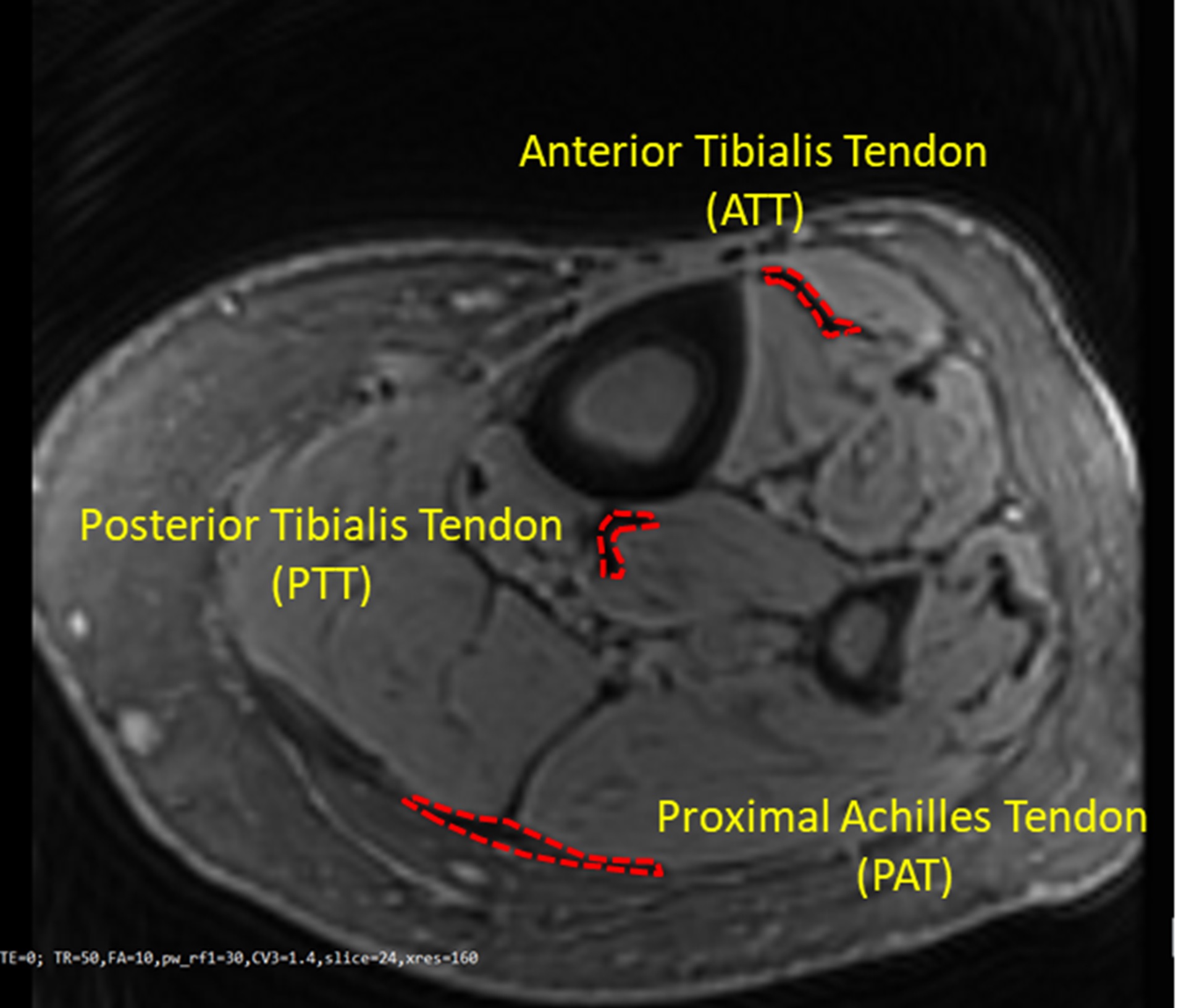
Figure 1: A
representative Cones UTE-MRI image from a 76-year-old female subject (TR=50 ms, for the selection the region of interests TE=2
ms was used because its provided higher contrast). Anterior
and posterior tibialis (ATT and PTT) and proximal Achilles (PAT) tendons
were obvious in the MRI images, as indicated in red.
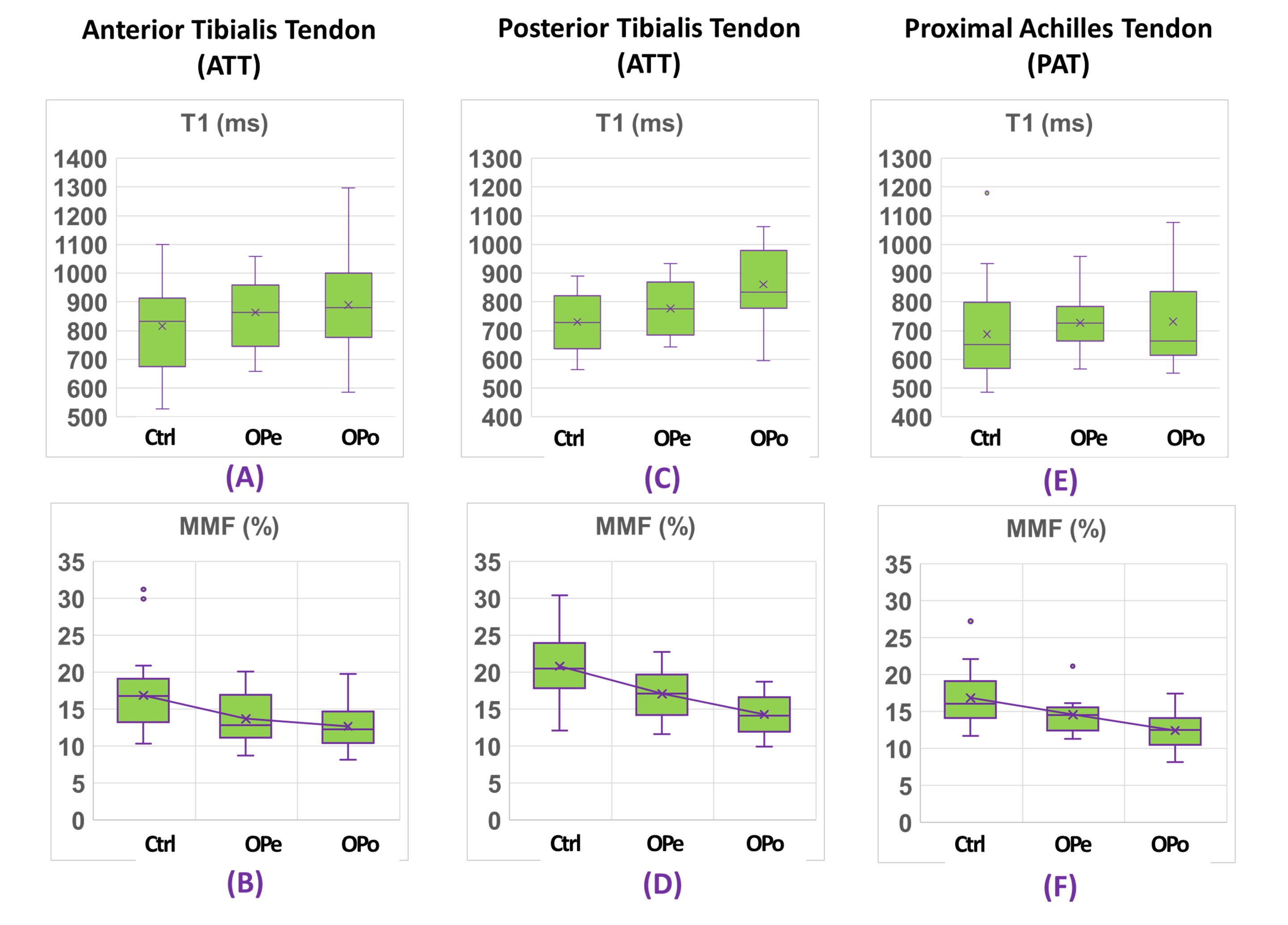
Figure
3: Boxplots of (A, C, E) T1 and (B, D, F) MMF of (A,B) ATT, (C,D) PTT, and
(E,F) PAT tendons in Ctrl subjects versus OPe and OPo patients. Averages,
median, SD, first, and third quartiles are indicated in the boxplots.