Ashley A. Williams1,2, Karyn E. Chappell1,2, and Constance R. Chu1,2
1Orthopaedic Surgery, Stanford University, Stanford, CA, United States, 2Veterans Affairs Palo Alto Health Care System, Palo Alto, CA, United States
1Orthopaedic Surgery, Stanford University, Stanford, CA, United States, 2Veterans Affairs Palo Alto Health Care System, Palo Alto, CA, United States
Meniscus and cartilage UTE-T2* was compared to arthroscopy
in degenerative meniscus tear patients.
Elevated UTE-T2* was observed in degenerate menisci. However, increasing
intra-operative cartilage grade was not associated
with strictly increasing cartilage UTE-T2*.
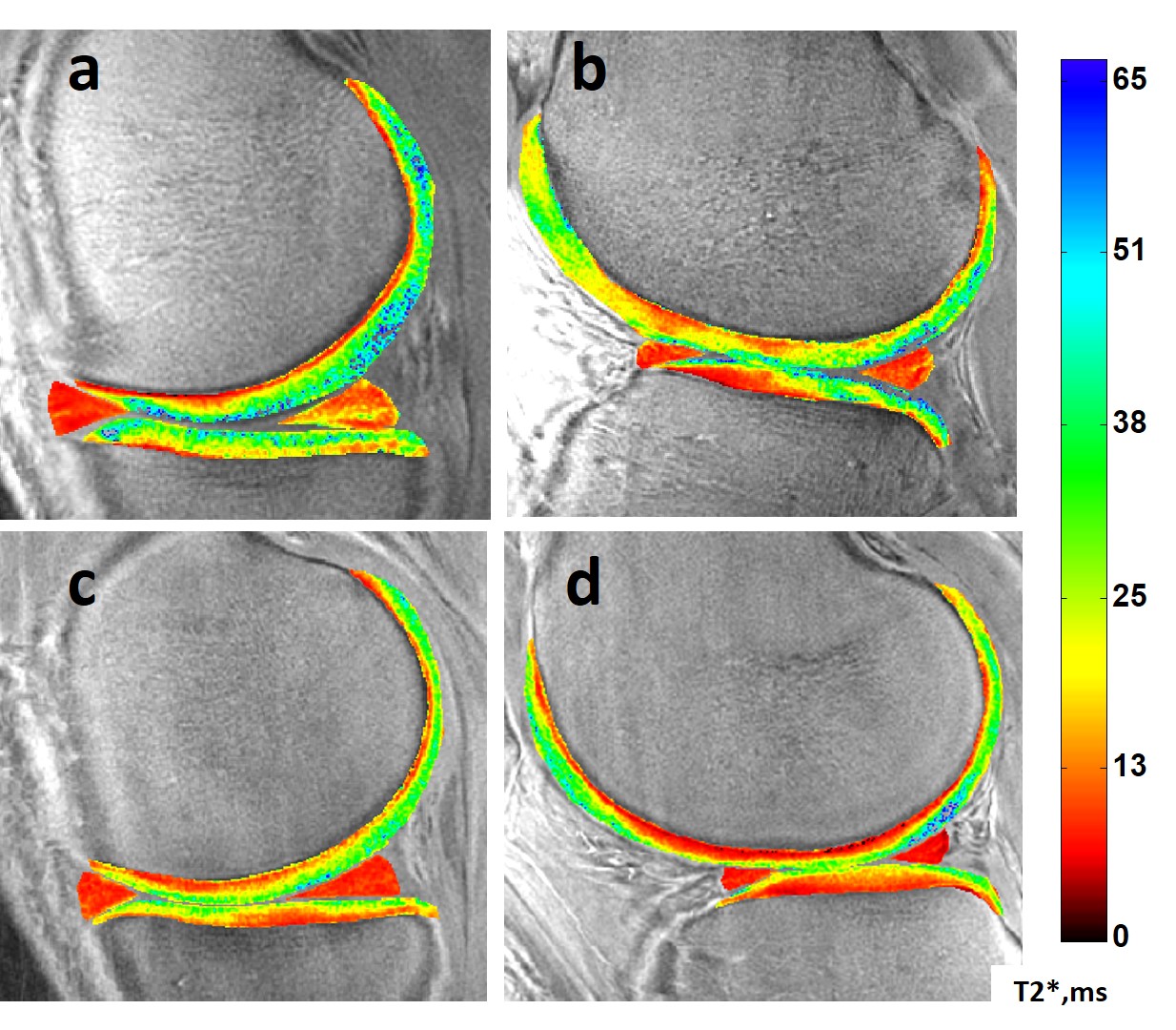
Figure 2. Sample UTE-T2* maps. Top row: a 39-yr male DMT patient with a complex tear
to the posterior horn of his medial meniscus (a) and arthroscopically detected
intact-but-softened medial and lateral femoral condylar cartilage but partial
to full-thickness disruptions of his tibial cartilages (a,b). Bottom row: an uninjured, healthy 24-yr male
with homogeneously low UTE-T2* in both medial and lateral menisci and smoothly
laminar UTE-T2* distributions in his articular cartilage (c,d).
Figure 1. Softened but intact cartilage regions (scope grade 1)
tend to have elevated deep cartilage UTE-T2* values compared to uninjured
controls (yellow bars, a,b,c), while cartilage regions with disrupted
articular surfaces tend to show UTE-T2* values consistent with or lower than
uninjured controls (red bars, a-d).
ANOVA (or Kruskal-Wallis) found no significant differences between
groups suggesting a high degree of variability in deep cartilage UTE-T2* values
of DMT patients with both intact and disrupted articular surfaces. Error bars
represent ±
standard error of the mean.