Nian Wang1, Anthony J. Mirando2, Yi Qi3, Matthew J. Hilton2, and Charles E. Spritzer3
1Radiology and Imaging Sciences, Indiana University, Indianapolis, IN, United States, 2Department of Orthopaedic Surgery, Duke University, Durham, NC, United States, 3Department of Radiology, Duke University, Durham, NC, United States
1Radiology and Imaging Sciences, Indiana University, Indianapolis, IN, United States, 2Department of Orthopaedic Surgery, Duke University, Durham, NC, United States, 3Department of Radiology, Duke University, Durham, NC, United States
FA decreased and MD increased after 8 weeks destabilization of the medial meniscus surgery.
Diffusion tractography affords a unique way to visualize the 3D collagen fiber architecture.
Diffusion MRI can be used as imaging biomarker for detecting the degradation of OA.

Figure 4. The fiber tracts of articular cartilage for both DMM (b) and SHAM (a) rats.
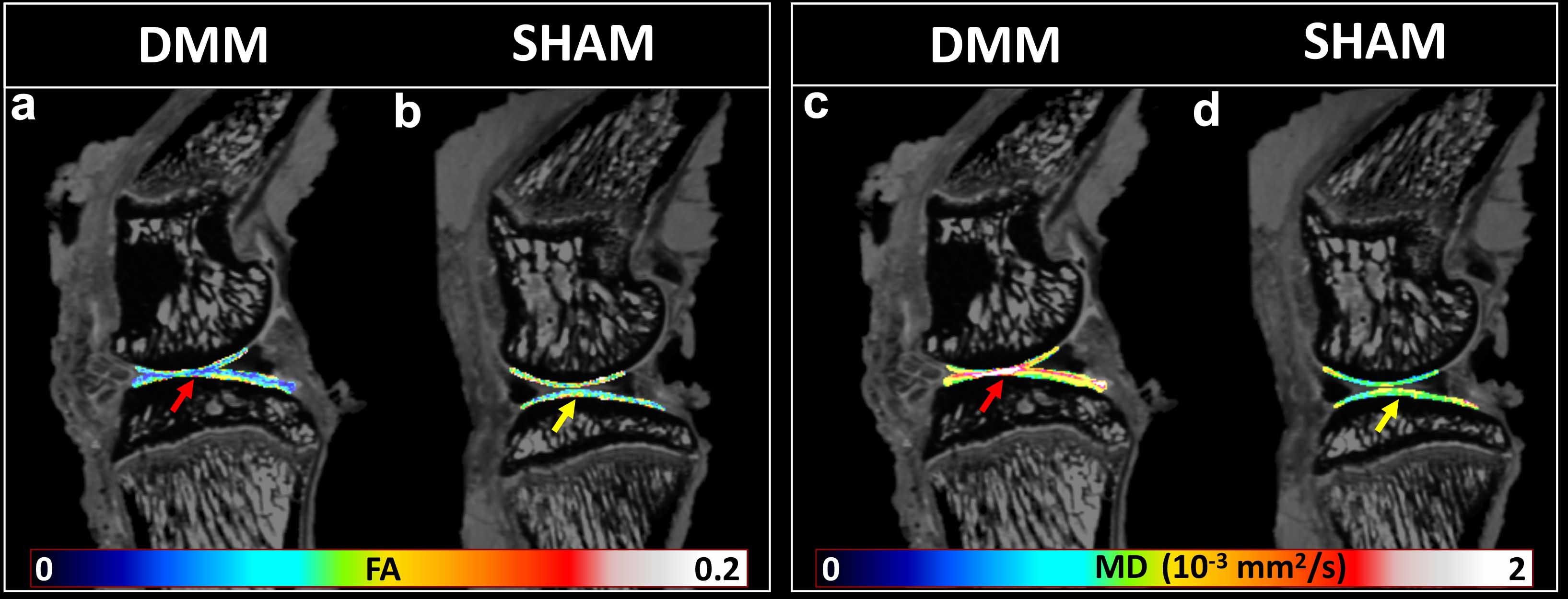
Figure 2. The quantitative fractional anisotropy (FA) and mean diffusivity (MD) maps of articular cartilage for DMM and SHAM rats.