Jose M Raya1, Alejandra M Duarte1, Dalibel M Bravo1, Elisa M Ramos1, Chongda M Zahng1, Mary M Cowman2, Thorsten M Kirsch1, Mark Wilne3, Len Lyut3, and Amparo M Ruiz1
1New York University Langone Health, New York, NY, United States, 2New York University Tandon School of Engineering, New York, NY, United States, 3University of Western Ontario, Ontario, ON, Canada
1New York University Langone Health, New York, NY, United States, 2New York University Tandon School of Engineering, New York, NY, United States, 3University of Western Ontario, Ontario, ON, Canada
This study is the first direct in vivo
indication for the involvement of inflammation in cartilage after injury. Our results provide
insights into the pathogenesis and role of inflammation in OA, showing that
hyaluronan-related inflammation is a common finding.
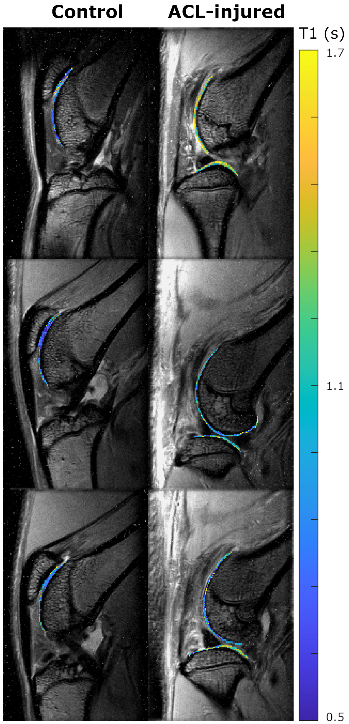
Figure 3. Example of T1 maps on control and ACL-injured limb before IA injection of Cy5.5-P15-1 (top row), and 24 h (middle row) and 48 h (bottom row) after injection. Control showed constant T1 values over time, while ACLT-injured limbs have a trend of decreased T1 values.
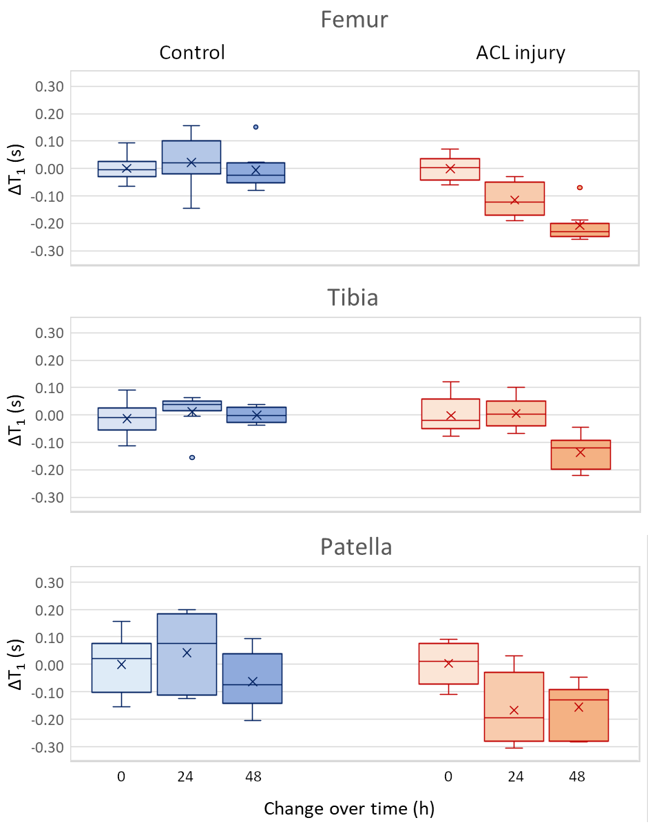
Figure 4. Boxplot showing the change of T1 values after injection of Cy5.5-P1-1 in controls (blue) and ACL-injured limbs (red) for femoral, tibial and patellar cartilage. Both femoral and patellar cartilage showed a significant difference in ΔT1 (p<0.05, one-way ANOVA).