Peiqi Ma1, Yushan Yuan1, Zongxi Zhang1, Bin Peng1, Jian Xu1, and Xiuzheng Yue2
1Fuyang People's Hospital,Anhui Province,China, Fuyang, China, 2Philips Healthcare, Beijing, China, Beijing, China
1Fuyang People's Hospital,Anhui Province,China, Fuyang, China, 2Philips Healthcare, Beijing, China, Beijing, China
In
this study, CS-SENSE in 3D acquisition for knee joint can not only improve the
scanning speed, but also more accurately diagnosis the type of meniscus injury.
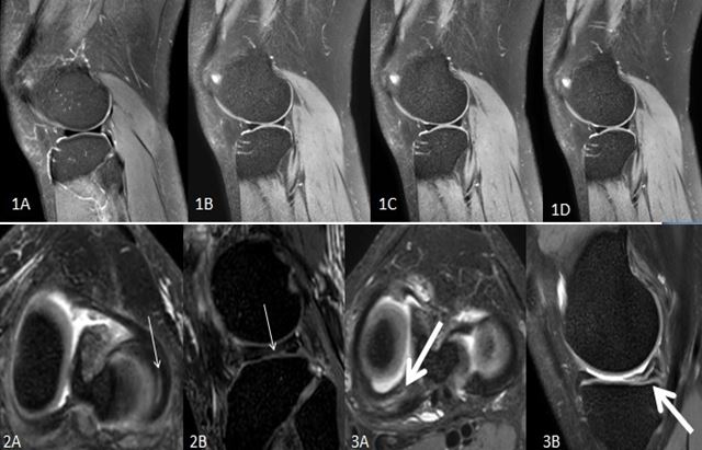
Figures 1A-1D show conventional 2D-MRI sagittal fsPDWI, CS-DSweak, CS-DSmediumand CS-DSstrongspectively.Figures 2A and 2B showlongitudinal cracks of the lateral meniscus(thin arrow). 2A is the reformatted transverse axis of CS-SENSE 3D scanning image (slicethickness of 1mm), and 2B is the reformatted sagittal images. Figures 3A and 3B show the horizontal crack in the posterior angle of the medial meniscus. 3A is the reformatted transverse axis of CS-SENSE 3D scanning image (slicethickness of1mm), and 3B is the reformatted sagittal images.