Shinji Saruya1, Masashi Suzuki1, Masami Yoneyama2, Kaiji Inoue1, Eito Kozawa1, and Mamoru Niitsu1
1Department of Radiology, Saitama Medical University, Saitama, Japan, 2Philips Japan, Tokyo, Japan
1Department of Radiology, Saitama Medical University, Saitama, Japan, 2Philips Japan, Tokyo, Japan
MIXTURE based on 3D multi-interleaved TSE offers rapid high-resolution isotropic T1rho mapping in the knee joint. Initial results in human subjects show promise for s improving the quality and efficiency of 3D T1ρ-mapping in the clinical practice.
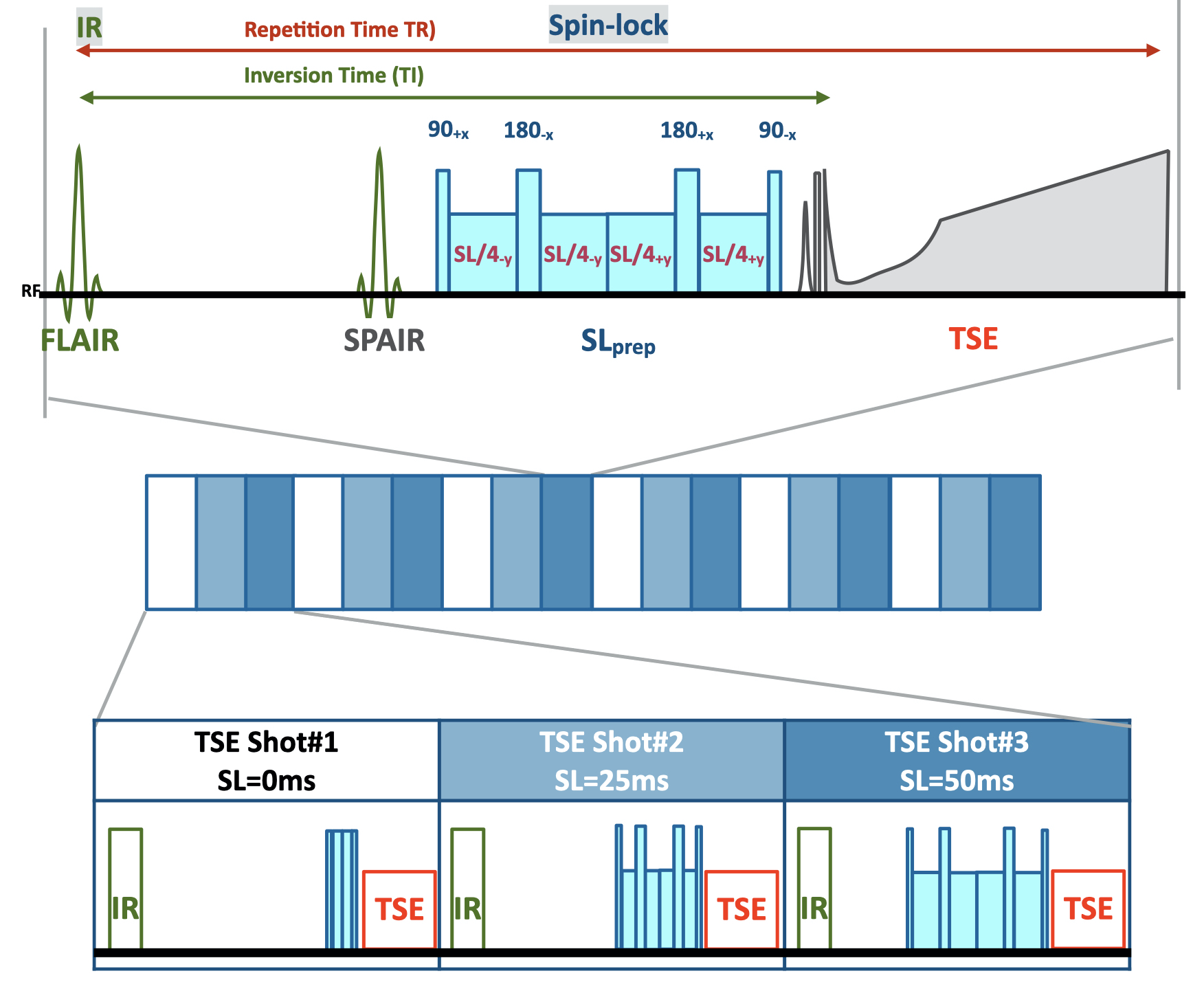
Figure 1. Scheme of the MIXTURE T1FLAIR T1ρ-mapping.T1ρ mapping was performed using an inversion-recovery and double-refocusing spin-lock prepared segmented TSE sequence with spectral fat suppression. Inversion-recovery was used for suppression of synovial fluid. Three images with different SL preparation times (SL = 0, 25, and 50ms) were acquired with interleaved acquisition. Amplitude of the SL pulse was set to 500 Hz.
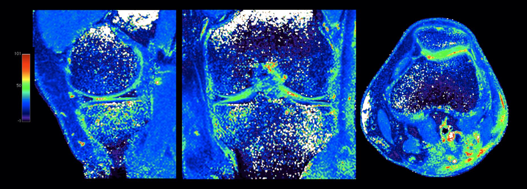
Figure 4. Representative sagittal, coronal, and axial MPR images of 3D isotropic T1ρ-mapping with 0.8mm3 acquisition using MIXTURE. The optimized sequence provided motion-insensitive high-quality isotropic images less than 9 minutes.