Jimin Ren1,2, Craig R Malloy1,2, Wanpen Vongpatanasin3, and Jarett Berry3
1Advanced Imaging Research Center, UT Southwestern Medical Center, Dallas, TX, United States, 2Department of Radiology, UT Southwestern Medical Center, Dallas, TX, United States, 3Department of Internal Medicine, UT Southwestern Medical Center, Dallas, TX, United States
1Advanced Imaging Research Center, UT Southwestern Medical Center, Dallas, TX, United States, 2Department of Radiology, UT Southwestern Medical Center, Dallas, TX, United States, 3Department of Internal Medicine, UT Southwestern Medical Center, Dallas, TX, United States
Pi accumulation and PCr depletion were linearly correlated during exercise. Post-exercise
ATP synthesis rate constant indexed by τ(Pi) and τ(PCr) were also linearly correlated in our elderly subjects (correlation coefficient 0.86, p-value =0.006, N = 8 subjects).
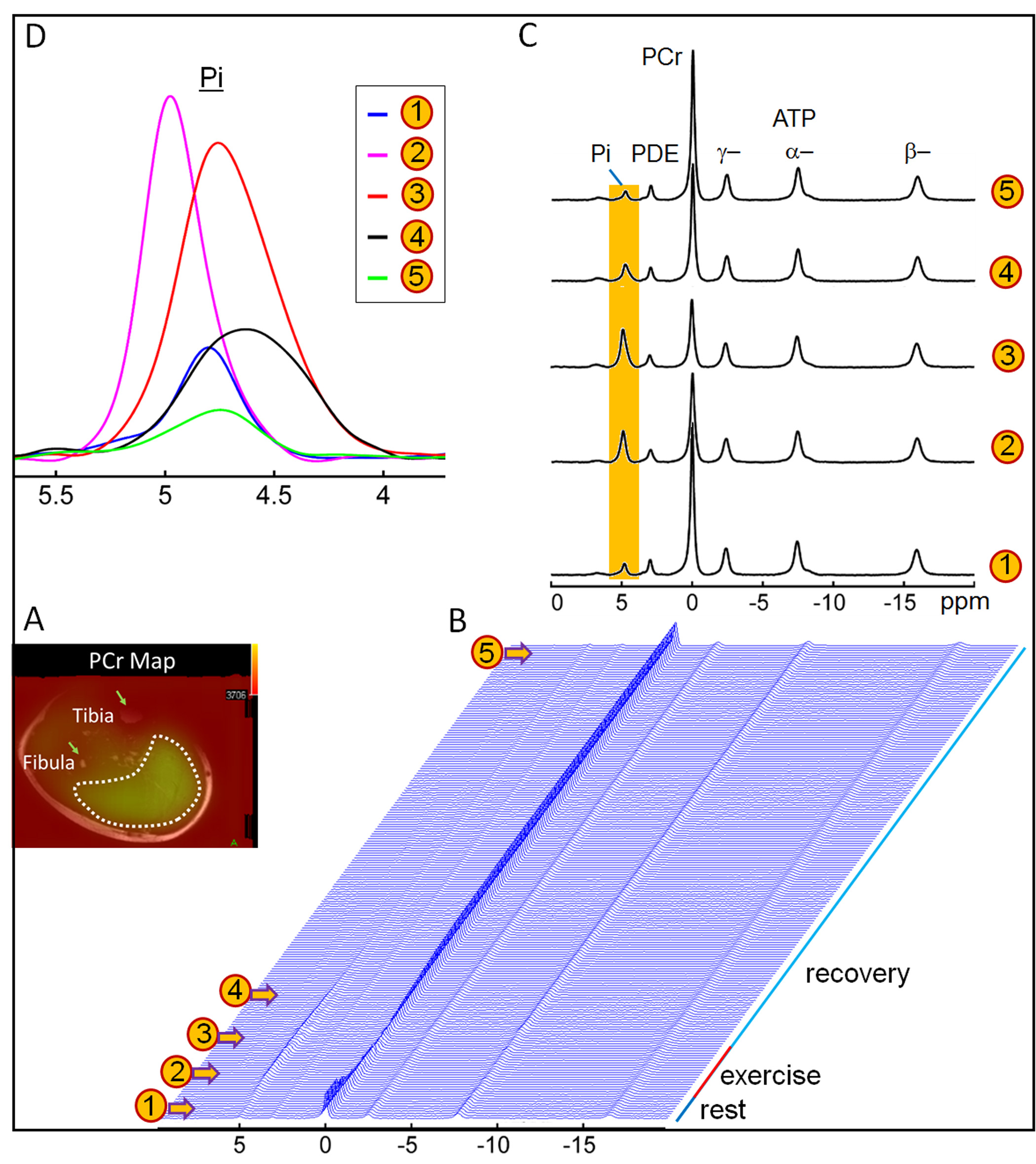
Fig.1 Dynamic
31P MR spectra acquired from calf muscle at 7T. (A) Cross-sectional T2W image showing coil
sensitive region located in calf muscle. (B) Dynamic 31P spectra at temporal
resolution of 2 s collected at rest, during exercise and recovery. (C)
Representative spectra at selected time points, with the highlighted Pi signals
expanded in (D).
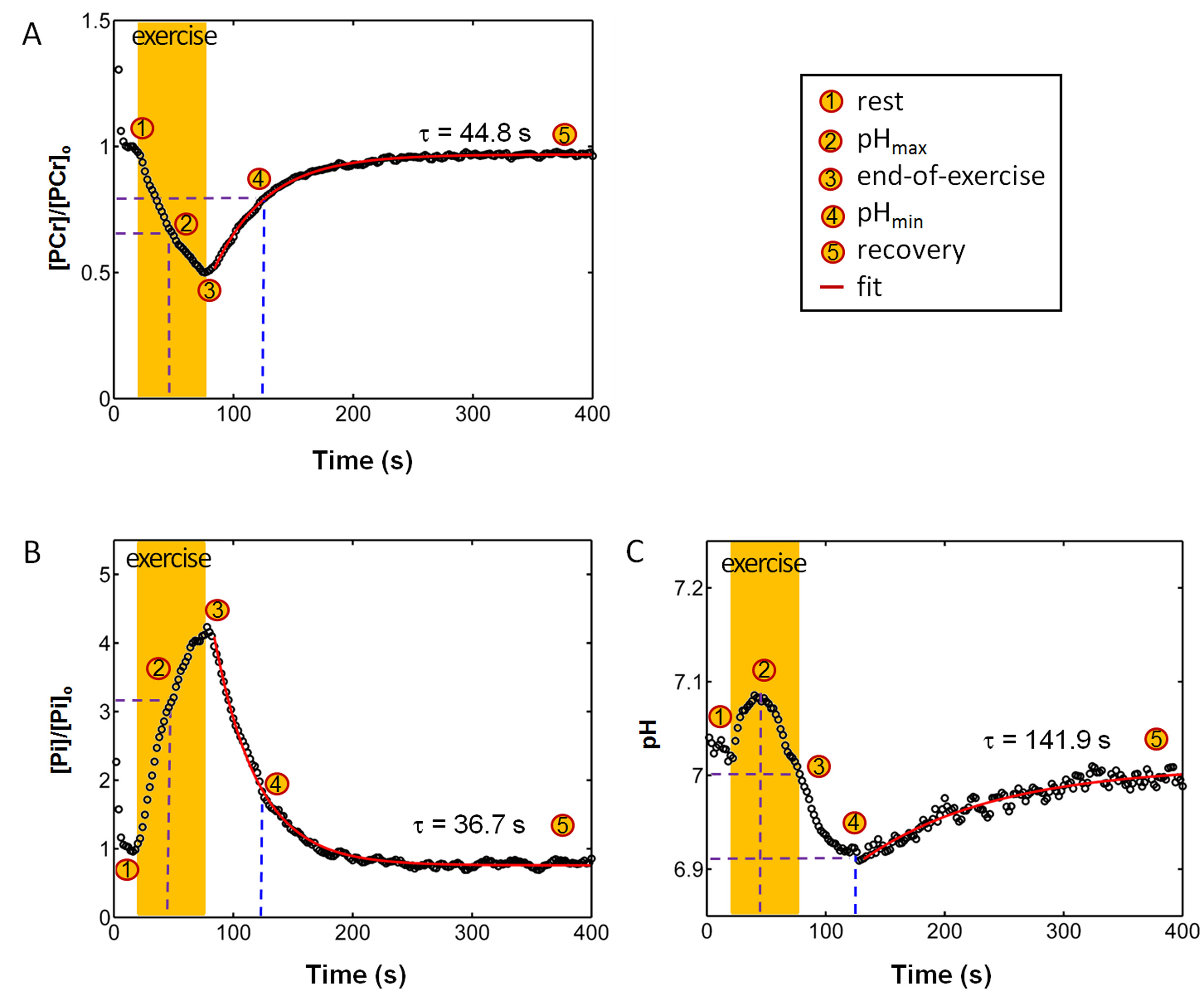
Fig.2 Plots
of time course of PCr depletion-recovery (A), Pi rise-decay (B), and pH (C). PCr
and Pi signals at resting steady-state are used as references. Note that the pH
plot clearly indicates the metabolic changes with alkalization in early
exercise and acidification in early recovery.