Takahiro Ueda1, Yoshiharu Ohno1,2, Kaori Yamamoto3, Kazuhiro Murayama2, Masato Ikedo3, Masao Yui3, Akiyoshi Iwase4, Takashi Fukuba4, Satomu Hanamatsu1, Yuki Obama1, Hirotaka Ikeda1, and Hiroshi Toyama1
1Radiology, Fujita Health University School of Medicine, Toyoake, Japan, 2Joint Research Laboratory of Advanced Medical Imaging, Fujita Health University School of Medicine, Toyoake, Japan, 3Canon Medical Systems Corporation, Otawara, Japan, 4Radiology, Fujita Health University Hospital, Toyoake, Japan
1Radiology, Fujita Health University School of Medicine, Toyoake, Japan, 2Joint Research Laboratory of Advanced Medical Imaging, Fujita Health University School of Medicine, Toyoake, Japan, 3Canon Medical Systems Corporation, Otawara, Japan, 4Radiology, Fujita Health University Hospital, Toyoake, Japan
DLR method is useful for improving image quality and
diagnostic performance of DWI without any adverse effect on ADC assessment
using a 3T MR system for patients with prostatic cancer.
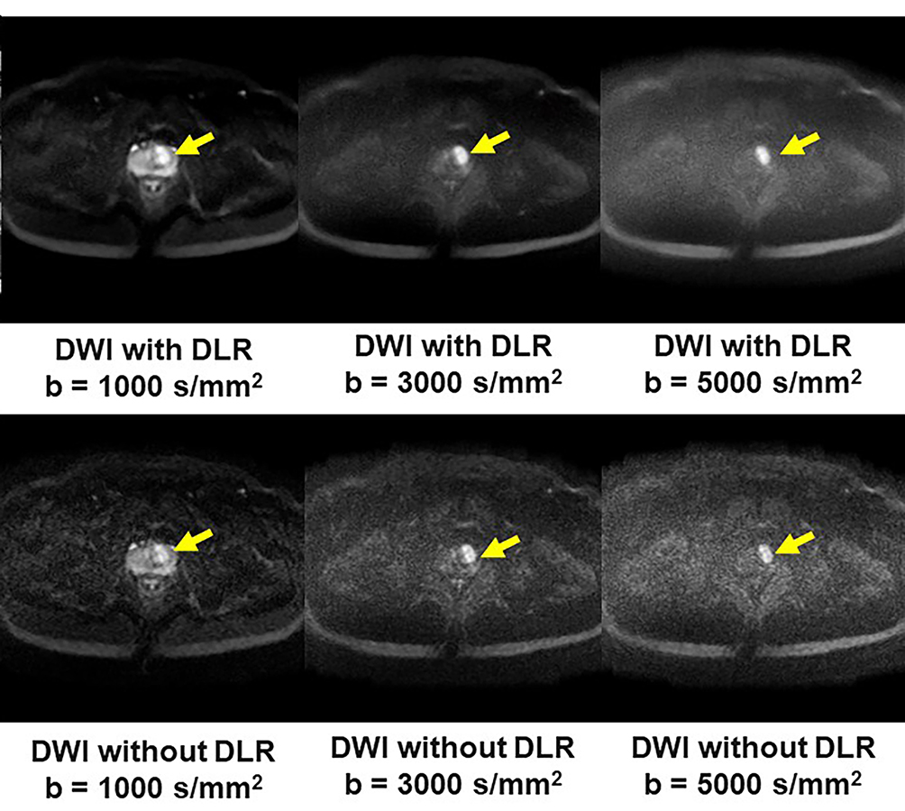
Figure 1. 51-year old patient with prostatic cancer in the left transitional zone
b: DLR improves image quality of DWIs for each b value. Each DWI shows the prostatic cancer as high signal intensity in the left transitional zone (arrow).
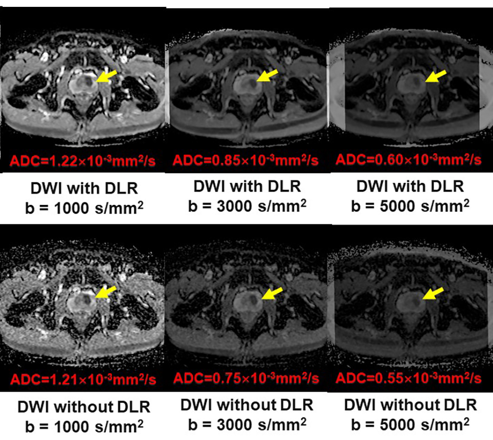
Figure 1. 51-year old patient with prostatic cancer in the left transitional zone
c: All ADC maps show the prostatic cancer as a hypointense signal in the left transitional zone (arrow).