Mohammed Saleh1, Sanaz Javadi1, Manoj Mathew2, Jong Bum Son3, Jia Sun4, Ersin Bayram5, Xinzeng Wang5, Jingfei Ma3, Janio Szklaruk1, and Priya Bhosale1
1Radiology, MD Anderson Cancer Center, Houston, TX, United States, 2Radiology, Stanford University, Stanford, CA, United States, 3Imaging Physics, MD Anderson Cancer Center, Houston, TX, United States, 4Biostatistics, MD Anderson Cancer Center, Houston, TX, United States, 5Global MR Applications and Workflow, GE Healthcare, Houston, TX, United States
1Radiology, MD Anderson Cancer Center, Houston, TX, United States, 2Radiology, Stanford University, Stanford, CA, United States, 3Imaging Physics, MD Anderson Cancer Center, Houston, TX, United States, 4Biostatistics, MD Anderson Cancer Center, Houston, TX, United States, 5Global MR Applications and Workflow, GE Healthcare, Houston, TX, United States
Compared to FSE, PROPELLER
showed reduced motion artifacts for T2-weighted imaging of pelvis. Deep Learning-based
image reconstruction (DL Recon) further improved image quality with better image
SNR, increased image sharpness and reduced artifacts.
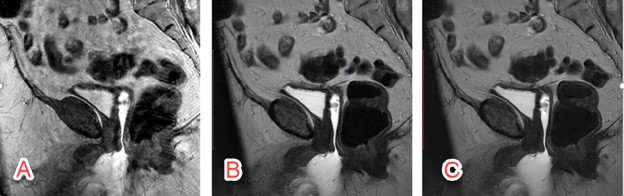
Figure 1. The conventional T2-weighted FSE images and PROPELLER images without and with
DL are shown. Because the phase encoding direction of AP was often chosen to avoid
aliasing and long scan time, conventional T2-weighted FSE is sensitive to
motion artifacts in sagittal imaging (A). PROPELLER is robust to motion and
minimized the motion artifacts (B), however, the SNR and in-plane resolution
were limited by the clinical scan time. DL reconstruction improved the SNR and
in-plane resolution without increasing scan time (C).
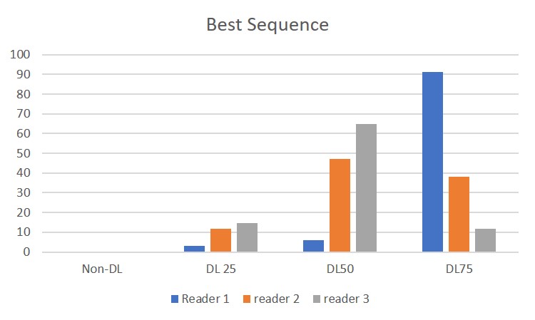
Figure 3. This graph shows the scoring of the best
sequence on the 4 images by three radiologists.
DL 50 and DL 75 were considered to be better than Non-DL and DL25.