Uma Sharma1, Vishwa Rawat1, Prasenjit Das 2, Achal Kumar Srivastava3, and Govind Makharia4
1Nuclear Magnetic Resonance and MRI Facility, All India Institute of Medical Sciences, New Delhi, India, 2Pathology, All India Institute of Medical Sciences, New Delhi, India, 3Neurology, All India Institute of Medical Sciences, New Delhi, India, 4Gasteroenterology and Human Nutrition, All India Institute of Medical Sciences, New Delhi, India
1Nuclear Magnetic Resonance and MRI Facility, All India Institute of Medical Sciences, New Delhi, India, 2Pathology, All India Institute of Medical Sciences, New Delhi, India, 3Neurology, All India Institute of Medical Sciences, New Delhi, India, 4Gasteroenterology and Human Nutrition, All India Institute of Medical Sciences, New Delhi, India
Volumetric analysis of
whole brain in gluten ataxia (GA) patients using MRI revealed significantly low brain and cerebellar
volumes along the lobules which form part of vermis while cerebrum volume is
not linked to GA.
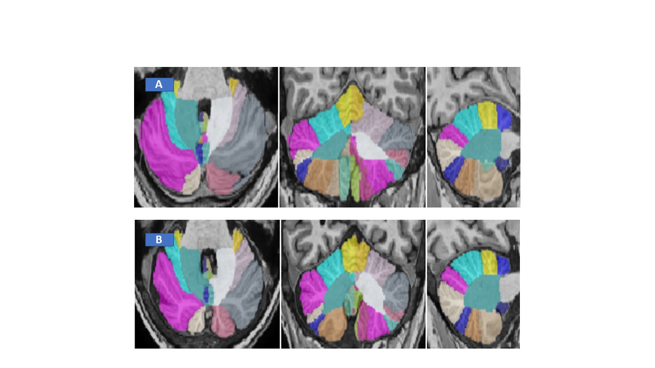
Figure 2: Representative CERES image of cerebellum taken from
Healthy control (A) and GA patient (B).
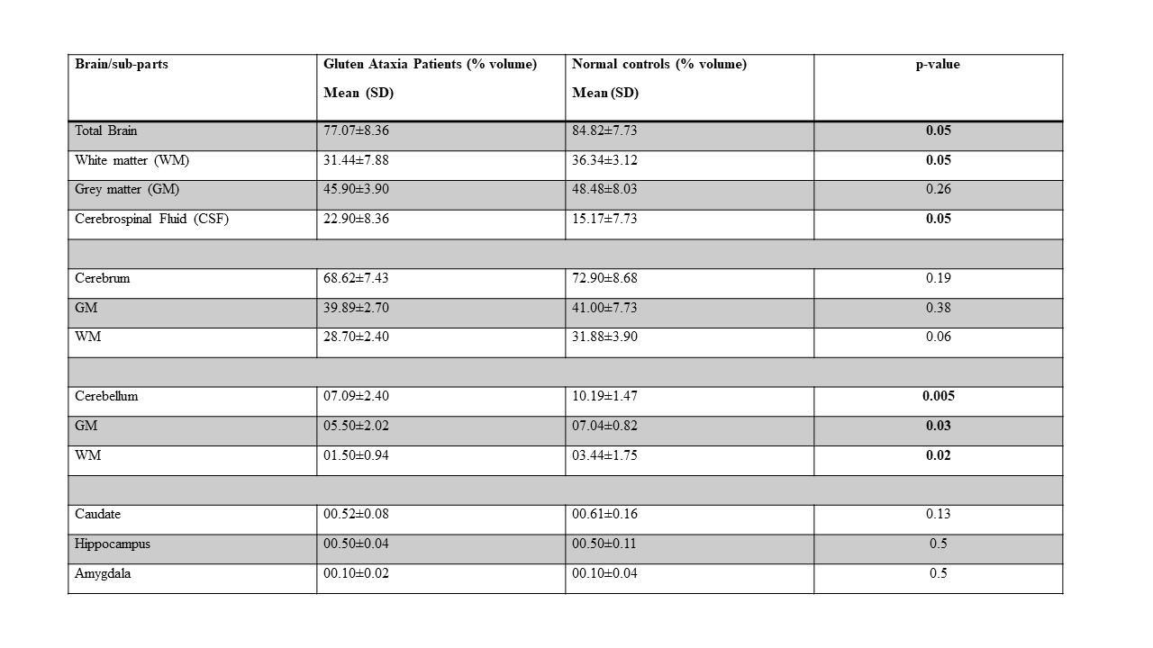
Table 2: Volumetric
estimation of Brain structures of GA patients and Healthy Controls.