Zenghui Cheng1, Bin Xiao2, Naying He3, Dinggang Shen4, Qian Wang5, Feng Shi4, Youmin Zhang3, Pei Huang3, Yan Li3, Sean K Sethi6, Kiarash Ghassaban7, Shengdi Chen3, Fuhua Yan3, and Ewart Mark Haacke7
1Radiology, Ruijin Hospital, Shanghai Jiao Tong University School of Medicine, Shanghai, China, 2Medical Imaging Technology, 、School of Biomedical Engineering, Shanghai Jiao Tong University, Shanghai, China, 3Ruijin Hospital, Shanghai Jiao Tong University School of Medicine, Shanghai, China, 4Shanghai United Imaging Intelligence Co., Ltd., Shanghai, China, 5Medical Imaging Technology, School of Biomedical Engineering, Shanghai Jiao Tong University, Shanghai, China, 6Magnetic Resonance Innovations, Inc, Bingham Farms, MI, United States, 7Wayne State University, Detroit, MI, United States
1Radiology, Ruijin Hospital, Shanghai Jiao Tong University School of Medicine, Shanghai, China, 2Medical Imaging Technology, 、School of Biomedical Engineering, Shanghai Jiao Tong University, Shanghai, China, 3Ruijin Hospital, Shanghai Jiao Tong University School of Medicine, Shanghai, China, 4Shanghai United Imaging Intelligence Co., Ltd., Shanghai, China, 5Medical Imaging Technology, School of Biomedical Engineering, Shanghai Jiao Tong University, Shanghai, China, 6Magnetic Resonance Innovations, Inc, Bingham Farms, MI, United States, 7Wayne State University, Detroit, MI, United States
Iron deposition and neuromelanin-containing neuron loss is prominent in the ventral and medial part of SNpc in early-stage PD. This region may correspond to nigrosome 2.
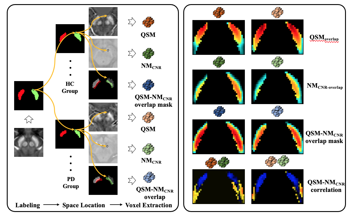
Figure 1. Flow of chart illustrating the voxel-wised analysis. PD- Parkinson’s disease, HC-healthy control, QSM- quantitative susceptibility mapping, NMCNR- contrast to noise of neuromelanin.
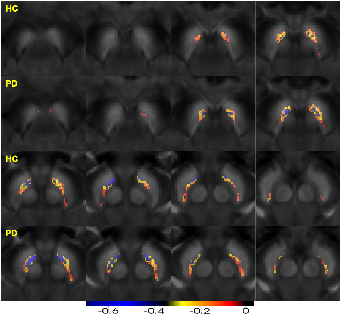
Figure 4. Negative Pearson’s correlation map of substantia nigra pars compacta (QSM-NMCNR overlap) PD- Parkinson’s disease, HC-healthy control, QSM- quantitative susceptibility mapping, NMCNR- contrast to noise of neuromelanin.