Septian Hartono1,2, An Sen Tan3, Weiling Lee4, Joey Oh4, Kuan Jin Lee5, Jongho Lee6, Eng King Tan1,2, and Ling Ling Chan2,4
1National Neuroscience Institute, Singapore, Singapore, 2Duke-NUS Medical School, Singapore, Singapore, 3Lee Kong Chian School of Medicine, Nanyang Technological University, Singapore, Singapore, 4Singapore General Hospital, Singapore, Singapore, 5Singapore BioImaging Consortium, Singapore, Singapore, 6Seoul National University, Seoul, Korea, Republic of
1National Neuroscience Institute, Singapore, Singapore, 2Duke-NUS Medical School, Singapore, Singapore, 3Lee Kong Chian School of Medicine, Nanyang Technological University, Singapore, Singapore, 4Singapore General Hospital, Singapore, Singapore, 5Singapore BioImaging Consortium, Singapore, Singapore, 6Seoul National University, Seoul, Korea, Republic of
Neuromelanin-sensitive MRI showed no significant differences between PD LRRK2 carriers and non-carriers. Quantitative susceptibility mapping was able to distinguish the two groups,
with higher substantia nigra (SN) iron deposition and larger high-iron area of the SN in PD LRRK2 carriers.
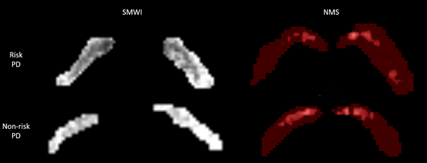
SN ROI derived from (1) semi-automated
segmentation on SMWI images (left) by thresholding for voxels with high iron
deposition containing signal intensity 7 standard deviations less than background
in and (2) NMS (right) by thresholding for voxels with signal intensity 4 standard
deviations higher than background in PD patient LRRK2 risk-variant
carriers (top row) and non-carriers (bottom row).
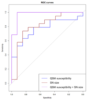
ROC analysis of QSM susceptibility, high-iron SN
size derived from SMWI, and combination model of QSM susceptibility and high-iron
SN size to classify LRRK2 risk-variant carriers and non-carriers in PD patients.