Jiahao Li1,2, Kelly Gillen1, Ilhami Kovanlikaya1, Thanh Nguyen1, Alexey Dimov1, Kailyn Li1, Weiyuan Huang1, Xianfu Luo1, Carly Skudin1, Eileen Chang1, Alexander Shtilbans1,3, and Yi Wang1,2
1Weill Cornell Medicine, New York, NY, United States, 2Meinig School of Biomedical Engineering, Cornell University, Ithaca, NY, United States, 3Hospital for Special Surgery, New York, NY, United States
1Weill Cornell Medicine, New York, NY, United States, 2Meinig School of Biomedical Engineering, Cornell University, Ithaca, NY, United States, 3Hospital for Special Surgery, New York, NY, United States
There
is an increase in susceptibility but a decrease in neuromelanin in the SN of PD
subjects as compared to controls.
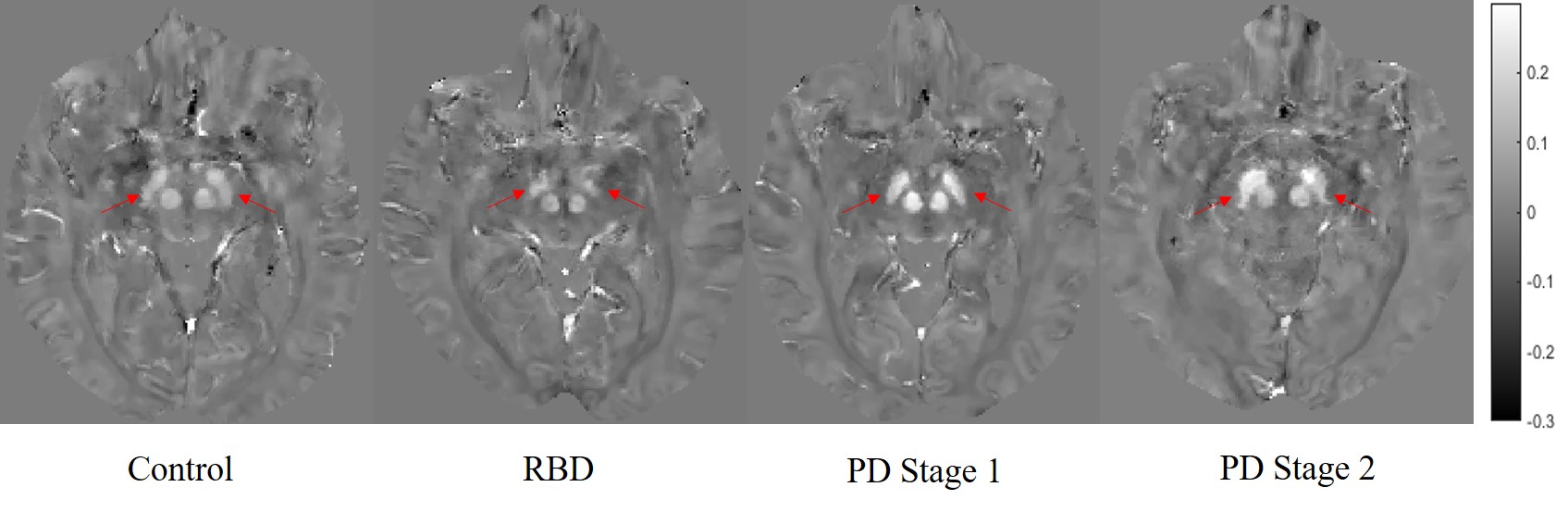
Figure
2. Representative QSM images from control, RBD, PD Stage 1 and PD Stage 2
subjects. Note increase
in susceptibility in substantia
nigra
(indicated
by red arrows) in PD
Stage 1 and
PD
Stage 2
compared to
healthy controls or RBD.
Scale bar in ppm (parts per million); RBD,
REM-sleep behavior disorder
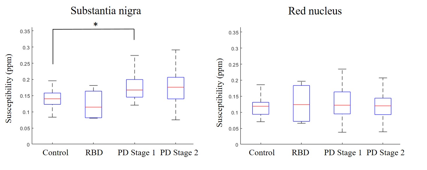
Figure
1. Average susceptibility in the substantia nigra
(SN; left) and red nucleus (RN; right) across all cohorts. Left: there is
significant increase in susceptibility PD
as compared
to controls. Right: there
are no statistically significant differences in susceptibility in the RN across
all cohorts. RBD,
REM-sleep behavior disorder; ppm, parts per million; *, p < 0.05 (multiple
comparison t-test, Bonferroni correction)