1Center for Mind/Brain Sciences - CIMeC, Mattarello, Italy
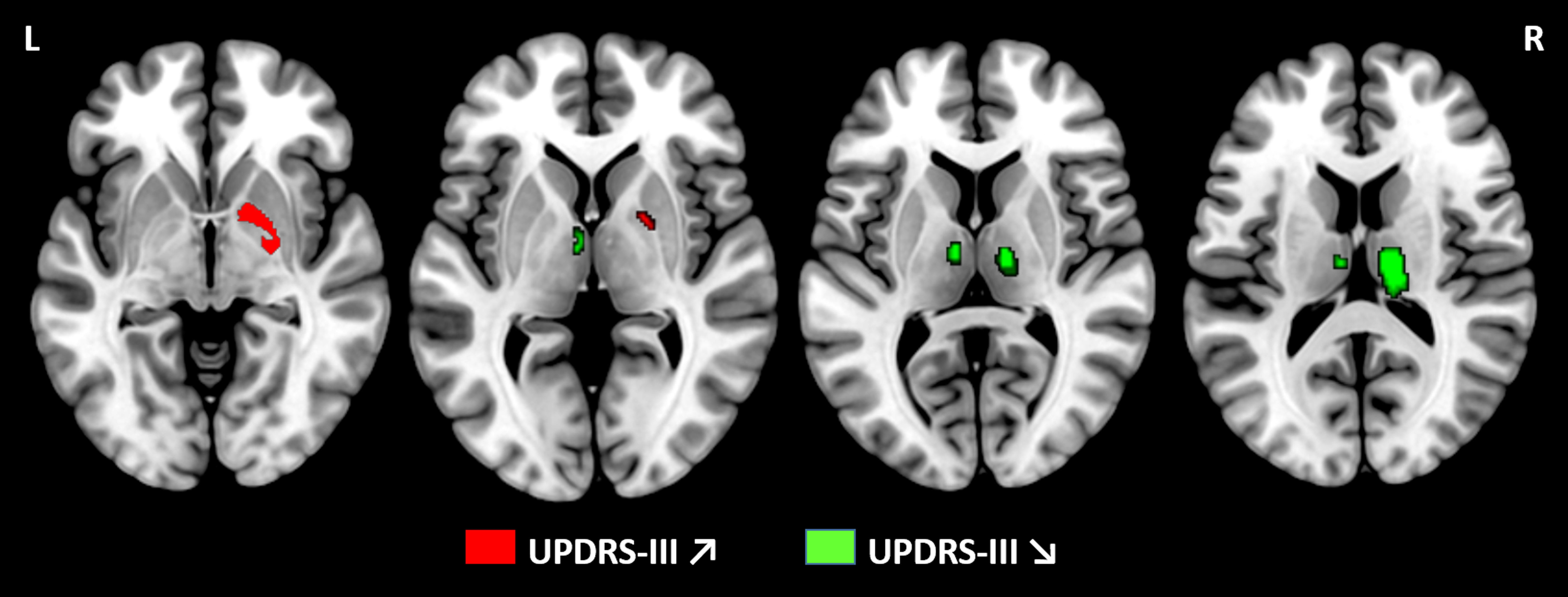
Figure 2: Statistical parametric maps showing positive and negative correlation between grey matter volume and UPDRS-III scores at 48 months using voxel-based morphometry analyses.
Results showed a positive association between grey matter volume and UPDRS-III in right pallidum at 48 months (p<0.05 TFCE, FWE corrected, age, sex and total intracranial volume adjusted). Conversely, a negative association was identified in bilateral thalamus at 48 months. No findings were significant at baseline.
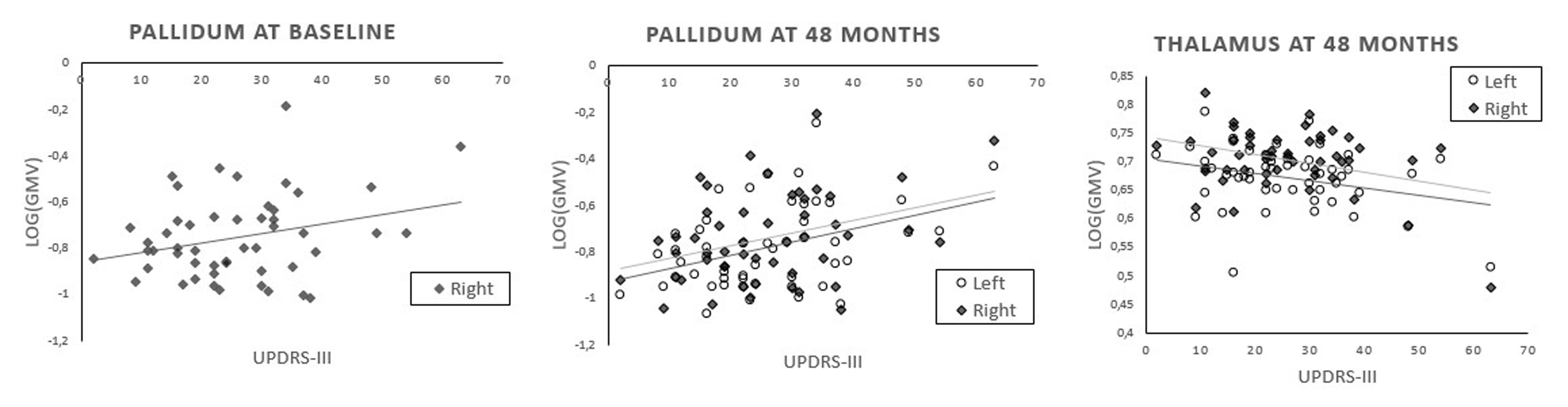
Figure 4: Correlation between grey matter volume and UPDRS-III scores at baseline and 48 months.
At baseline, one positive correlation was identified between grey matter volume and UPDRS-III scores in the right pallidum. At 48 months, a positive correlation was observed between grey matter volume and UPDRS-III scores in bilateral pallidum whereas a negative correlation was identified in bilateral thalamus. Pearson correlations are reported with a statistical threshold of p<0.05.