Chao Zhang1, Weiqiang Dou2, and Kai Xu1
1Department of Radiology, Xuzhou Medical University, Xuzhou, China, 2GE Healthcare,MR Research China, Beijing, China
1Department of Radiology, Xuzhou Medical University, Xuzhou, China, 2GE Healthcare,MR Research China, Beijing, China
Our study provides new
insights into the neural basis of cognitive dysfunction in PD patients. We also
found that BNM-FC can be an effective feature to distinguish PD-MCI from PD-NC.
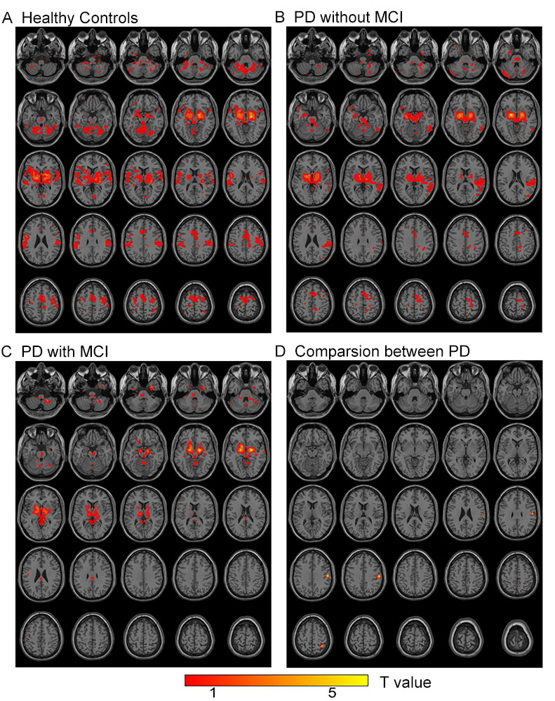
Figure 1. Functional
Connectivity (FC) between the basal nucleus of Meynert (BNM) and whole brain.
(A–C): BNM-FC was evaluated in healthy control (HC) (A), Parkinson’s disease with
normal cognition (PD-NC) (B), and Parkinson’s disease with mild cognitive impairment
(PD-MCI) (C) groups. (D): BNM-FC was decreased in the right superior parietal
lobe (SPL) of the PD-MCI group compared to the PD-NC and HC groups.
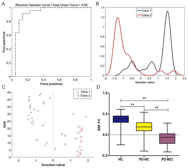
Figure 2.
Classification accuracy of functional connectivity (FC) in the basal nucleus of
Meynert (BNM) (BNM-FC) in the right superior parietal lobe (SPL) determined by
the leave-one-out cross-validation (LOOCV) method in distinguishing Parkinson’s
disease with mild cognitive impairment (PD-MCI) from Parkinson’s disease with
normal cognition (PD-NC) patient (A–C). Box plots with whiskers (min–max) show
BNM-FC in the right SPL of the three groups (D).