Yan Bai1,2, Rushi Chen1,2, Wei Wei1,2, Rui Zhang1,2, Zhun Huang2,3, Xianchang Zhang4, Mathias Nittka5, Gregor Koerzdoerfer5, and Meiyun Wang1,2
1Department of Radiology, Zhengzhou University People’s Hospital & Henan Provincial People’s Hospital, Academy of Medical Sciences., Zhengzhou, China, 2Henan Key Laboratory for Medical Imaging of Neurological Diseases, Zhengzhou, Henan, China, ZhengZhou, China, 3Department of Radiology, Henan University People’s Hospital & Henan Provincial People’s Hospital, School of Basic Medicine., Zhengzhou, China, 4MR Collaboration, Siemens Healthcare Ltd., Beijing, China, BeiJing, China, 5MR Pre-development, Siemens Healthcare GmbH, Erlangen, Germany, Erlangen, Germany
1Department of Radiology, Zhengzhou University People’s Hospital & Henan Provincial People’s Hospital, Academy of Medical Sciences., Zhengzhou, China, 2Henan Key Laboratory for Medical Imaging of Neurological Diseases, Zhengzhou, Henan, China, ZhengZhou, China, 3Department of Radiology, Henan University People’s Hospital & Henan Provincial People’s Hospital, School of Basic Medicine., Zhengzhou, China, 4MR Collaboration, Siemens Healthcare Ltd., Beijing, China, BeiJing, China, 5MR Pre-development, Siemens Healthcare GmbH, Erlangen, Germany, Erlangen, Germany
Conventional
magnetic resonance imaging cannot
reliably differentiate Parkinson’s disease (PD) from essential tremors (ET). Magnetic resonance fingerprinting (MRF) can simultaneously acquire T1 and
T2 relaxometry. This study utilized MRF to obtain T1 and T2 values in substantia
nigra (SN) of patients with tremor-dominant PD and ET. The T1 values of SN were
significantly higher in patients with tremor-dominant PD than those with ET, whereas
the T2 values showed no significant differences between groups. The findings
suggest that MRF T1 mapping of the SN can potentially differentiate tremor-dominant PD from ET
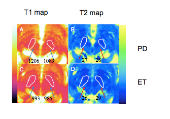
Figure 1. (A) T1 map and
(B) T2 map from a patient with tremor-dominant Parkinson’s disease (PD). (C) T1
map and (D) T2 map from a patient with essential tremors (ET). The T1 value in
the substantia nigra is significantly higher in the patient with
tremor-dominant PD than in the patient with ET.