Yu Shen1, Xianchang Zhang2, Yan Bai1, Yaping Wu1, Ewart Mark Haacke3, Bo Wu4, Yongsheng Chen4, Rui Zhang1, Rushi Chen1, Wei Wei1, and Meiyun Wang1
1Department of Medical Imaging, Henan Provincial People’s Hospital & Zhengzhou University, Zhengzhou, China, 2MR Collaboration, Siemens Healthcare Ltd. Beijing China, Beijing, China, 3Wayne State University, Detroit, MI, United States, 4Shanghai Zhuyan Medical Technology Company, Shanghai, China
1Department of Medical Imaging, Henan Provincial People’s Hospital & Zhengzhou University, Zhengzhou, China, 2MR Collaboration, Siemens Healthcare Ltd. Beijing China, Beijing, China, 3Wayne State University, Detroit, MI, United States, 4Shanghai Zhuyan Medical Technology Company, Shanghai, China
Strategically acquired gradient echo imaging is a useful and accurate method to examine structural brain changes and iron accumulation in patients with Parkinson’s disease.
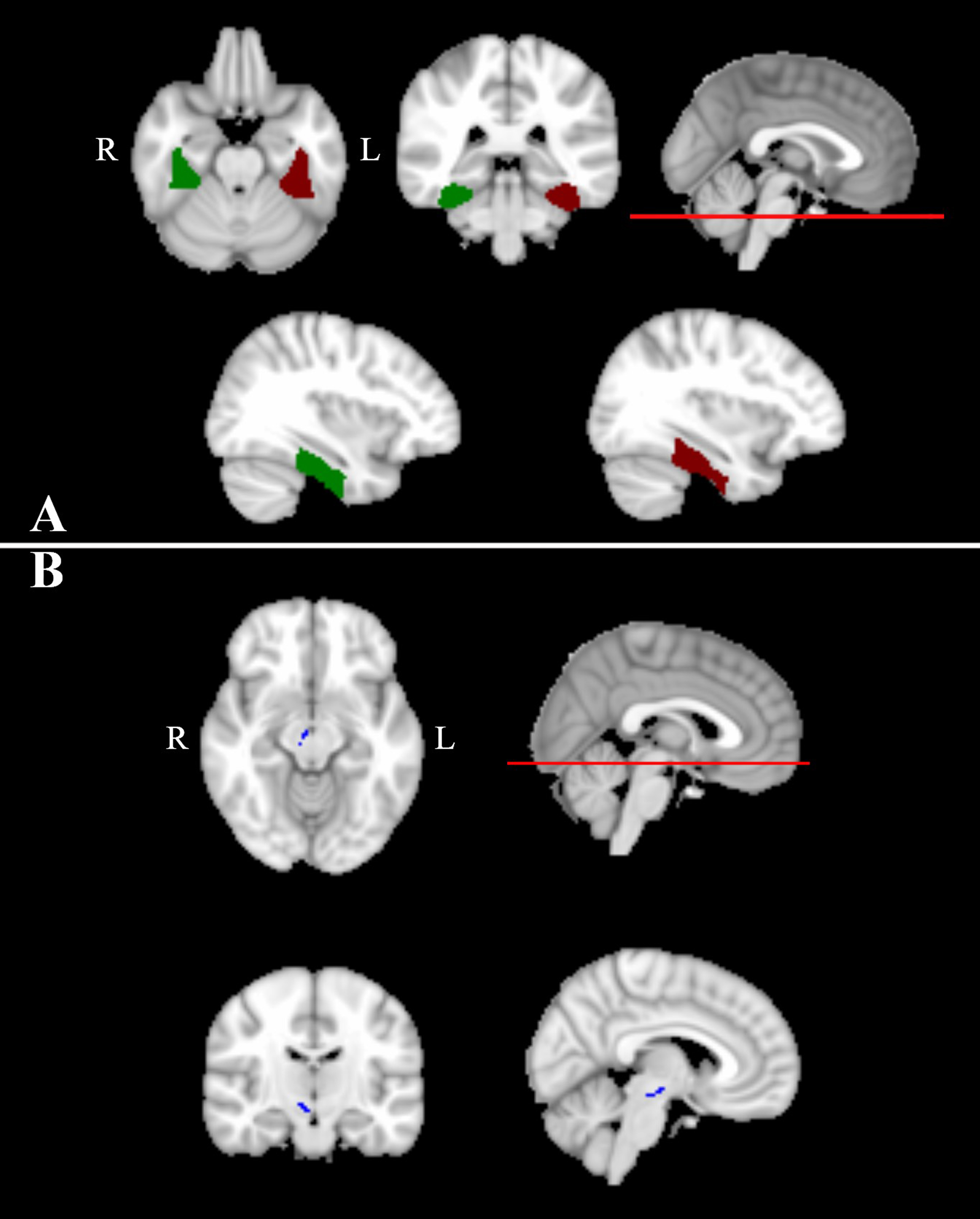
Figure 2. Images showing brain regions with significant differences between PD and healthy controls (HC). (A) The left temporal fusiform cortex, posterior division (TFCp) is shown in red, right TFCp is shown in green. (B) The right parabrachial pigmented (PBP) nucleus is shown in blue. R2* of right TFCp and right PBP was significantly higher in PD than in HC; T2* of left TFCp, right TFCp and right PBP was significantly lower in PD than in HC; T1 mapping ofright PBP was significantly lower in PD than in HC.
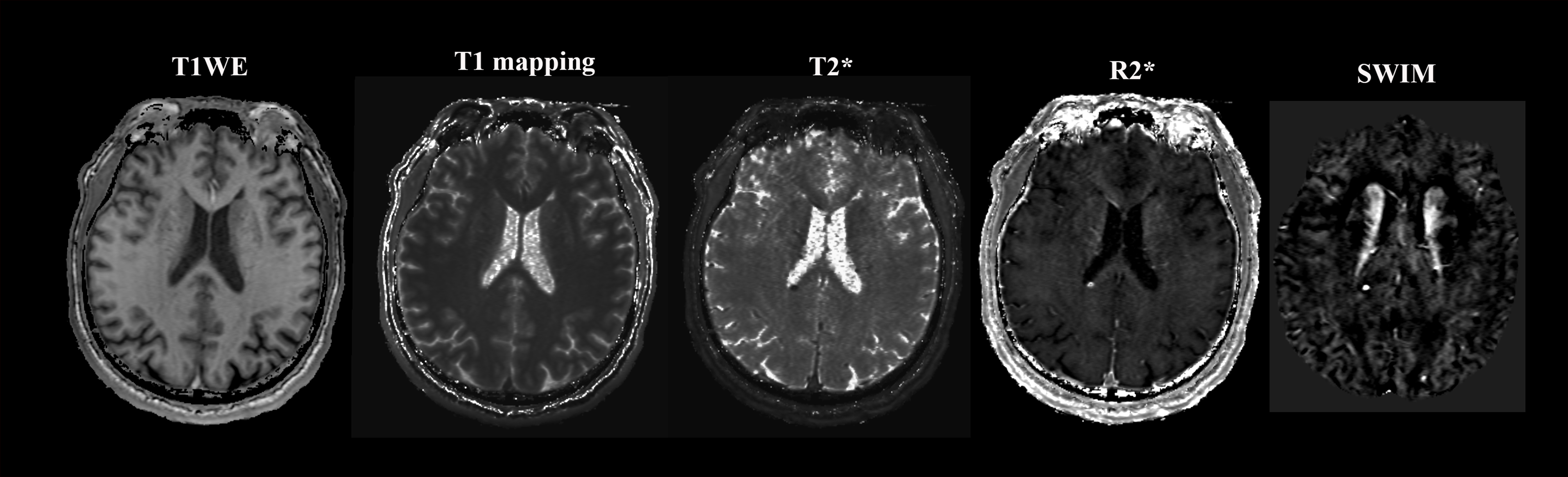
Figure 1. Images from T1-weighted enhanced imaging (T1WE), T1 mapping, T2*, R2*, and susceptibility-weighted imaging (SWI) mapping acquired using strategically acquired gradient echo imaging. T1WE shows improved image quality over the other methods.