1Department of Radiology, Juntendo University Graduate School of Medicine, Tokyo, Japan, 2Department of Radiological Sciences, Graduate School of Human Health Sciences, Tokyo Metropolitan University, Tokyo, Japan, 3Department of Radiology, Toho University Omori Medical Center, Tokyo, Japan
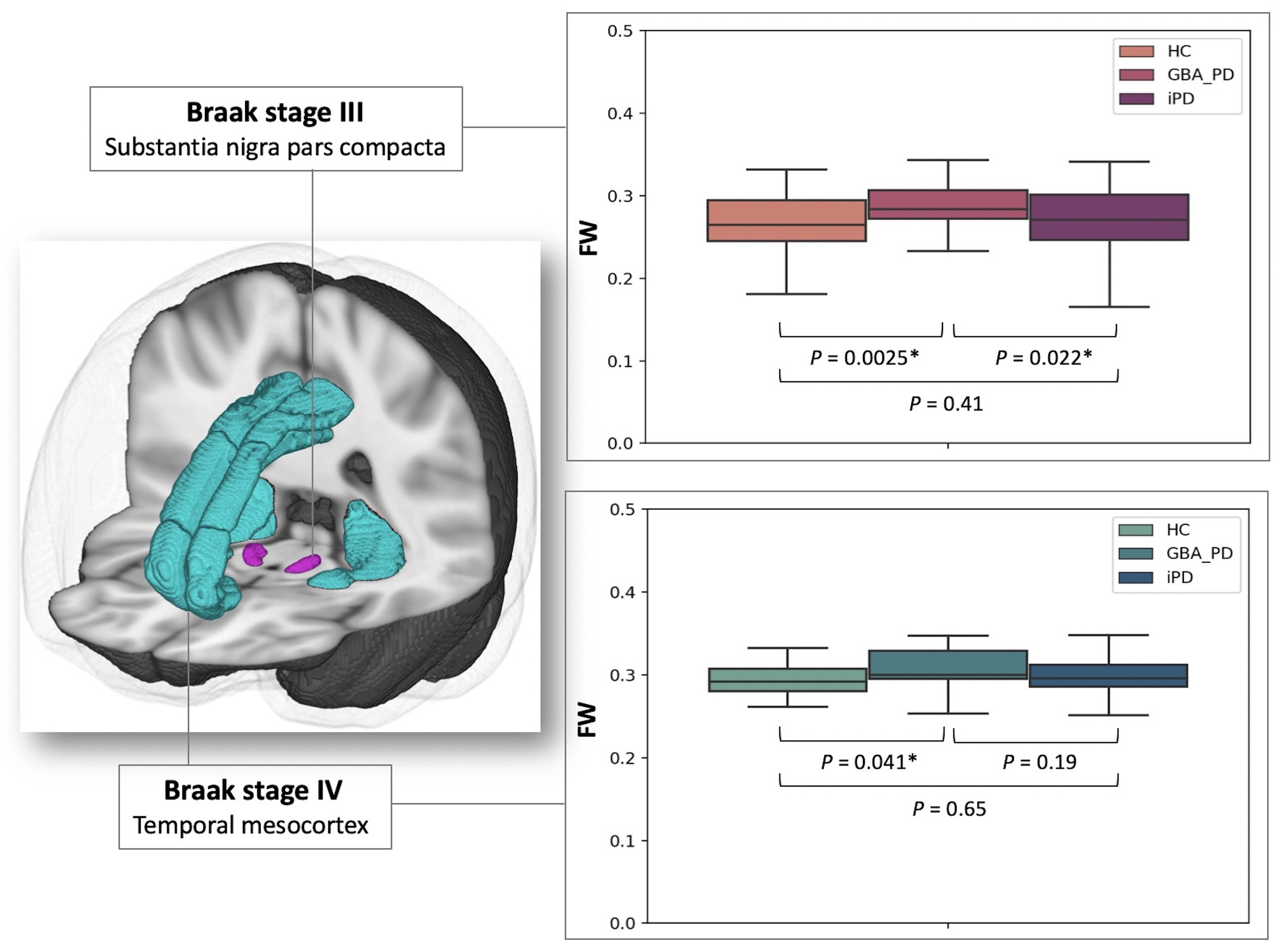
Figure 3. Box plots of FW for the HC, GBA-PD, and iPD groups in Braak stages III and IV. The bottom and top of the box are the first and third quartiles, and the thick band inside the box is the median. Whiskers represent maximum and minimum values of all data.
Abbreviations: FW, free water, GBA-PD, Parkinson’s disease with GBA1 mutations; HC, healthy controls; and iPD, idiopathic Parkinson’s disease.
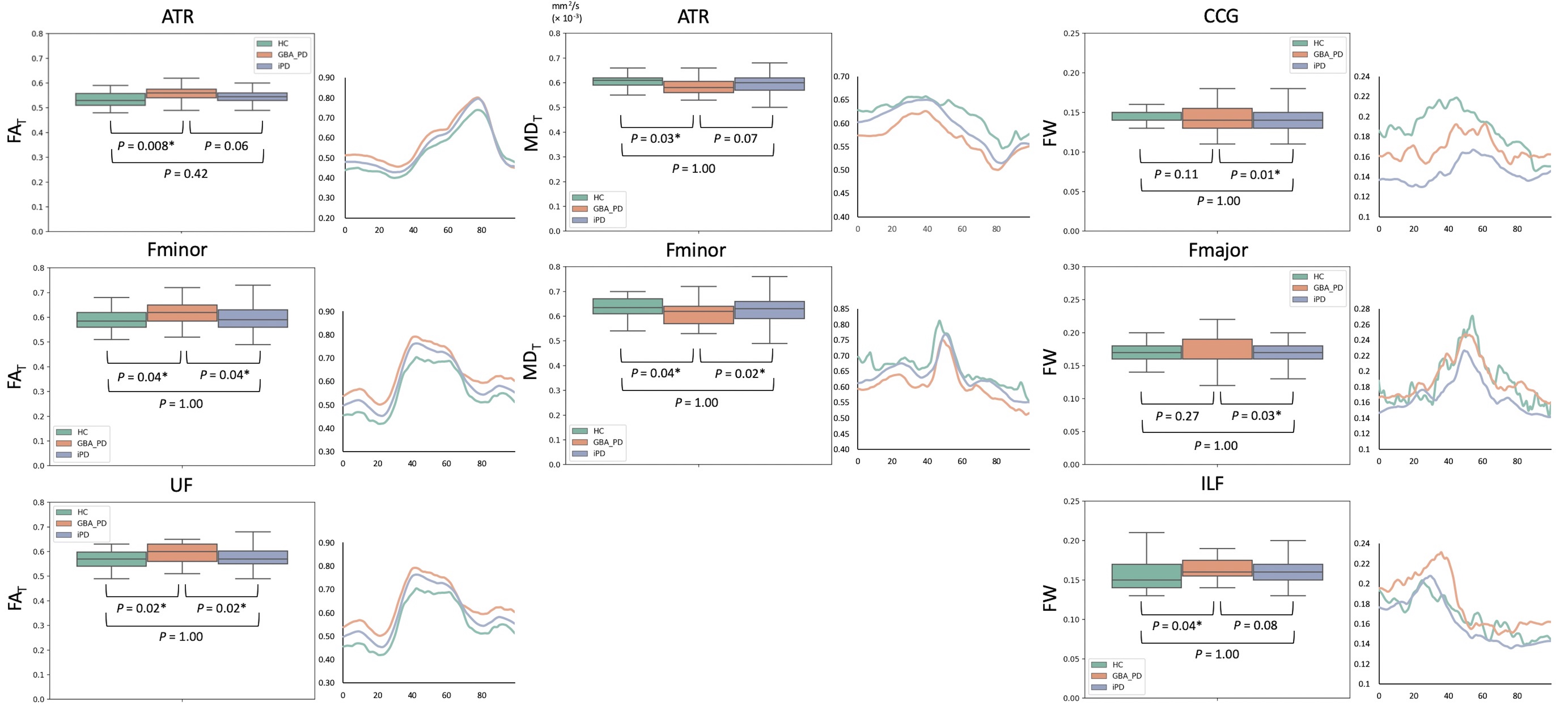
Figure 2. Box plots and tract profiles of FAT, MDT, and FW for HC, GBA-PD, and iPD groups in the white matter pathways.
Abbreviations: ATR, anterior thalamic radiation; CCG, cingulum cingulate gyrus; FAT, free water-corrected fractional anisotropy; Fmajor, forceps major; Fminor, forceps minor; FW, free water; GBA-PD, Parkinson’s disease with GBA1 mutations; HC, healthy controls; ILF, inferior longitudinal fasciculus; iPD, idiopathic Parkinson’s disease; MDT, free water-corrected mean diffusivity; UF, uncinate fasciculus.