1Advanced Clinical Imaging Technology, Siemens Healthcare AG, Bern, Switzerland, 2Translational Imaging Center, sitem-insel AG, Bern, Switzerland, 3Departments of Radiology and Biomedical Research, University of Bern, Bern, Switzerland, 4Siemens Healthcare GmbH, Erlangen, Switzerland, 5Advanced Clinical Imaging Technology, Siemens Healthcare AG, Lausanne, Switzerland, 6Department of Radiology, University Hospital (CHUV) and University of Lausanne (UNIL), Lausanne, Switzerland, 7LTS5, École Polytechnique Fédérale de Lausanne, Lausanne, Switzerland
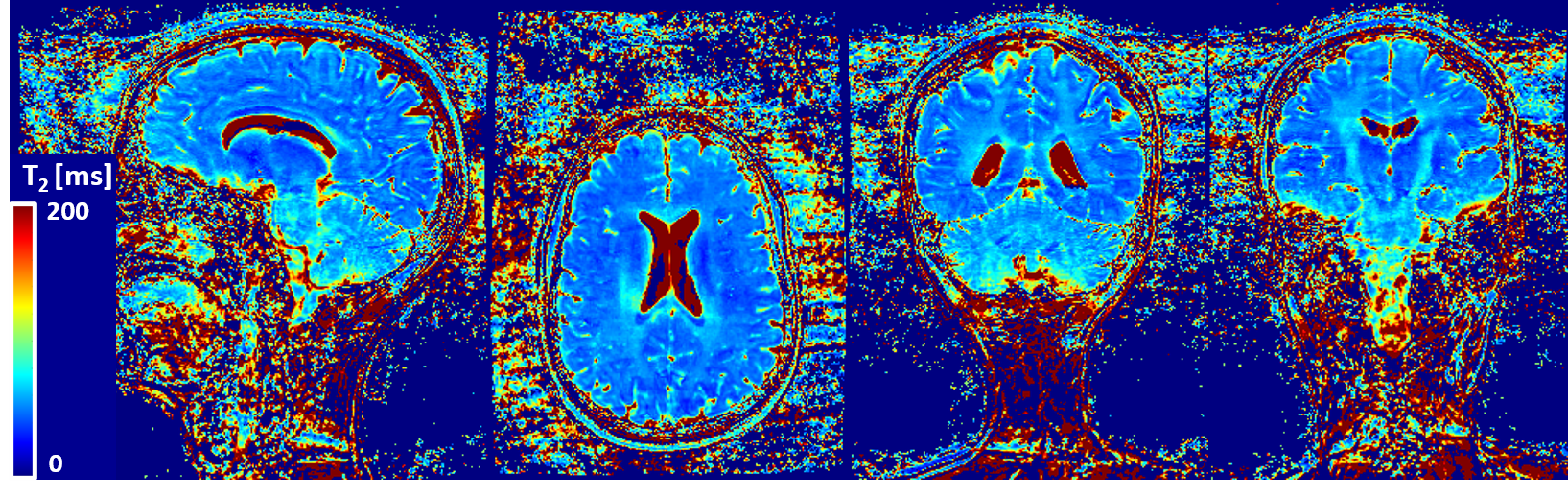
Figure 4
T2 map generated from T2-prepared volumes shown in figure 3. T2 values show good contrast between gray matter and white matter structures. T2 estimation may be compromised in the inferior portion of the cerebellum which may be due to B1 inhomogeneity.

Figure 3
Orthogonal views of T2-prepared images obtained from a healthy subject show good contrast and increasing T2 weighting as a function of T2-prep time. Fair signal homogeneity can be observed till the basal area of the brain with a slight signal drop in the cerebellum. High level of detail can be observed in the cerebellum and corpus callosum in the coronal views as benefit from sub-millimetric resolution. Signal-shift artifacts originating from cerebrospinal fluid (arrows) can be observed and will be investigated.